GLI2 anticorps (Middle Region)
 (23 références)
 (2 validations)
-
- Antigène Voir toutes GLI2 Anticorps
- GLI2 (GLI Family Zinc Finger 2 (GLI2))
-
Épitope
- Middle Region
-
Reactivité
- Humain
-
Hôte
- Lapin
-
Clonalité
- Polyclonal
-
Conjugué
- Cet anticorp GLI2 est non-conjugé
-
Application
- Western Blotting (WB), Immunohistochemistry (IHC)
- Séquence
- RNDVHLRTPL LKENGDSEAG TEPGGPESTE ASSTSQAVED CLHVRAIKTE
- Homologie
- Human: 100%
- Attributs du produit
- This is a rabbit polyclonal antibody against GLI2. It was validated on Western Blot and immunohistochemistry.
- Purification
- Protein A purified
- Immunogène
- The immunogen is a synthetic peptide directed towards the N terminal region of human GLI2
-
anti-GLI Family Zinc Finger 2 (GLI2) (AA 1287-1321), (C-Term) antibody
GLI2 Reactivité: Humain WB, IHC (p), IF, FACS Hôte: Lapin Polyclonal RB49147 unconjugated
anti-GLI Family Zinc Finger 2 (GLI2) (Internal Region) antibodyGLI2 Reactivité: Souris WB, ELISA, IHC Hôte: Lapin Polyclonal unconjugated
anti-GLI Family Zinc Finger 2 (GLI2) (Internal Region) antibodyGLI2 Reactivité: Humain, Chimpanzé WB, ELISA, IHC Hôte: Lapin Polyclonal unconjugated
anti-GLI Family Zinc Finger 2 (GLI2) (C-Term) antibodyGLI2 Reactivité: Humain WB, ELISA, IF, FACS Hôte: Lapin Polyclonal unconjugated
anti-GLI Family Zinc Finger 2 (GLI2) antibodyGLI2 Reactivité: Humain, Souris, Rat IF Hôte: Lapin Polyclonal unconjugated
anti-GLI Family Zinc Finger 2 (GLI2) (AA 600-649) antibodyGLI2 Reactivité: Humain WB, IHC, IHC (p) Hôte: Lapin Polyclonal unconjugated
anti-GLI Family Zinc Finger 2 (GLI2) (AA 2-20) antibodyGLI2 Reactivité: Humain WB, ELISA, IHC Hôte: Lapin Polyclonal unconjugated
anti-GLI Family Zinc Finger 2 (GLI2) (N-Term) antibodyGLI2 Reactivité: Humain, Souris, Rat WB, ELISA, IHC, IF, ICC Hôte: Lapin Polyclonal unconjugated
anti-GLI Family Zinc Finger 2 (GLI2) (AA 1444-1463), (C-Term) antibodyGLI2 Reactivité: Humain WB Hôte: Lapin Polyclonal unconjugated
anti-GLI Family Zinc Finger 2 (GLI2) antibodyGLI2 Reactivité: Souris, Rat WB Hôte: Lapin Polyclonal unconjugated
-
- Indications d'application
- Optimal working dilutions should be determined experimentally by the investigator.
- Commentaires
-
Antigen size: 1258 AA
- Restrictions
- For Research Use only
-
- by
- Laboratory of Herditary Cancer, Division of Molecular Medicine, Rudjer Boskovic Institute
- No.
- #101239
- Date
- 05.10.2017
- Antigène
- GLI2
- Numéro du lot
- QC49260-42551
- Application validée
- Western Blotting
- Contrôle positif
Human melanoma cell line WM793B
Human metastatic melanoma tissue
- Contrôle négative
- Human healthy skin not expressing GLI2
- Conclusion
- Passed. ABIN2777474 specifically recognizes GLI2 in WM793B cells and human metastatic melanoma tissue.
- Anticorps primaire
- ABIN2777474
- Anticorps secondaire
- anti-rabbit IgG HRP-linked antibody (Cell Signaling Technology, 7074)
- Full Protocol
- Grow WM793B cells in RPMI (Lonza, BE12-702F) supplemented with FBS (Sigma-Aldrich, F7524) and penicillin/streptomycin (Thermo Fisher Scientific, 15070063), at 37°C and 5% CO2 in a 10 cm TC-dish.
- Wash cells 2x with PBS.
- Harvest cells.
- Lyse cells in 85µl cold lysis buffer (25mM Tris-HCl pH7.5, 150mM NaCl, 1% NP-40, 1% sodium deoxycholate, 0.1% SDS) with sonication 2x 15s using an Labsonic M sonicator with a 1mm probe (B. Braun Biotech International, 8535027), 1 cycle, 80% amplitude.
- Determine total protein content of the lysates using a BCA protein assay (Thermo Fisher Scientific, 23225).
- Denature 50μg of total proteins for 5min at 95°C in Laemmli SDS sample buffer and subsequently separate them on a freshly cast denaturing 7% PAA gel (using components from Sigma-Aldrich) for 2h at 100V.
- Transfer proteins onto Amersham Protran Premium 0.2 NC membrane (GE Healthcare, 10600004) with a Mini Trans-Blot cell (Bio-Rad, 170-3930) for 80min at 100V.
- Block the membrane with blocking buffer and blocking reagent (TBST/5% non-fat milk) for 30min at RT.
- Incubation with primary rabbit anti human GLI2 antibody (antibodies-online, ABIN2777474, lot QC492960-42551) diluted 1:1000 in TBST/5% non-fat milk ON at 4°C.
- Wash membrane 3x for 5min with TBST.
- Incubation with secondary anti-rabbit IgG HRP-linked antibody (Cell Signaling Technology, 7074) diluted 1:3000 in 5% non-fat milk/TBST for 1h at RT.
- Wash membrane 3x for 5mins with TBST.
- Reveal protein bands using Super Signal West Pico and Super Signal West Femto reagents (Thermo Fisher Scientific) on an UVITEC Alliance Imaging system (Cambridge).
- Notes
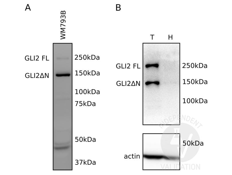
The GLI2 antibody ABIN2777474 reveals two protein bands of the expected molecular weight for the full-length (GLI2 FL) and the GLI2 N-terminally truncated (GLI2ΔN) isoform in lysates of WM793B cells.
ABIN2777474 also detects both GLI2 FL as well as GLI2ΔN in metastatic melanoma cell lysates whereas neither protein is revealed in human healthy skin tissue in which expression of GLI2 is not to be expected.
The predicted molecular weight of the full length GLI2 is 197kDa which is in accordance with our observation. With our previous anti-GLI2 antibody (sc-20291 (G-20), Santa Cruz Biotechnology) we also detected the GLI2ΔN protein at ~150kDa. The difference in apparent MW could be due to different migration of the marker.
Validation #101239 (Western Blotting)
 Validation Images
![A Detection of endogenously expressed GLI2 with ABIN2777474 in total cell lysates of the human melanoma cell line WM793B. Both the full length protein (GLI2 FL) as well as the ΔN (GLI2ΔN) isoform are detected. B Detection of endogenously expressed GLI2 with ABIN2777474 in human metastatic melanoma tissue (T) compared to human healthy skin tissue that does not express GLI2 (H). Both the full length protein as well as the ΔN isoform are only detected in metastatic melanoma.]() A Detection of endogenously expressed GLI2 with ABIN2777474 in total cell lysates of the human melanoma cell line WM793B. Both the full length protein (GLI2 FL) as well as the ΔN (GLI2ΔN) isoform are detected. B Detection of endogenously expressed GLI2 with ABIN2777474 in human metastatic melanoma tissue (T) compared to human healthy skin tissue that does not express GLI2 (H). Both the full length protein as well as the ΔN isoform are only detected in metastatic melanoma.
A Detection of endogenously expressed GLI2 with ABIN2777474 in total cell lysates of the human melanoma cell line WM793B. Both the full length protein (GLI2 FL) as well as the ΔN (GLI2ΔN) isoform are detected. B Detection of endogenously expressed GLI2 with ABIN2777474 in human metastatic melanoma tissue (T) compared to human healthy skin tissue that does not express GLI2 (H). Both the full length protein as well as the ΔN isoform are only detected in metastatic melanoma.
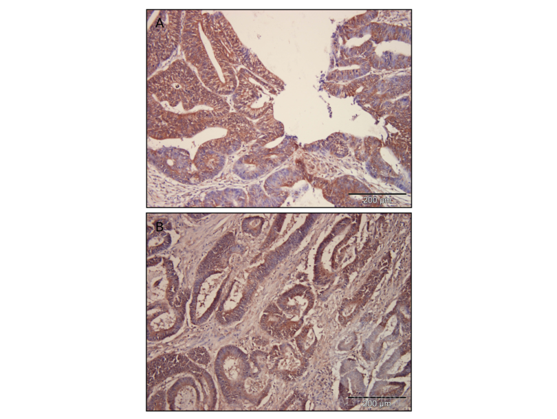
![Immunohistochemical staining GLI2 with ABIN2777474 in well differentiated colorectal carcinoma (A) and poorly differentiated colorectal carcinoma (B). Localization of GLI2 is cytoplasmic and nuclear. Magnification 200x.]() Immunohistochemical staining GLI2 with ABIN2777474 in well differentiated colorectal carcinoma (A) and poorly differentiated colorectal carcinoma (B). Localization of GLI2 is cytoplasmic and nuclear. Magnification 200x.
Protocole
Immunohistochemical staining GLI2 with ABIN2777474 in well differentiated colorectal carcinoma (A) and poorly differentiated colorectal carcinoma (B). Localization of GLI2 is cytoplasmic and nuclear. Magnification 200x.
Protocole -
- by
- Laboratory for Hereditary Cancer, Division of Molecular Medicine, Rudjer Boskovic Institute
- No.
- #102749
- Date
- 02.02.2018
- Antigène
- GLI2
- Numéro du lot
- QC49260-42551
- Application validée
- Immunohistochemistry
- Contrôle positif
- Human colon cancer tissue sections
- Contrôle négative
- no primary antibody
- Conclusion
Passed. ABIN2777474 specifically labels the targeted antigen in human colon cancer samples in IHC.
- Anticorps primaire
- ABIN2777474
- Anticorps secondaire
- biotinylated anti-rabbit, streptavidin-HRP (LSAB2 System-HRP, Dako, K0675, lot 10126805)
- Full Protocol
- Fix human colon cancer tissue in 10% buffered formalin, ON at RT.
- Process tissue using an automated tissue processor (Leica or Tissue-Tec VIP Sakura):
- 70% ethanol 2x for 30min.
- 96% ethanol 2x for 30min.
- Absolute ethanol 2x 30min.
- Xylene substitute (Tissue-Clear, Sakura) 2x 30min.
- Paraffin 2x 30min at 62°C. Embed tissue in paraffin blocks using the Tissue-Tec TEC embedding console system.
- Cut paraffin embedded tissue with a Sliding microtome Slide 4003 E (PFM Medical) into 3µm sections.
- Deparaffinize and rehydrate sections through a graded xylene and graded alcohol series:
- Bioclear New xylene substitute (Biognost, BCN-1L, lot BCN-03/17) 3x for 3min.
- 100% ethanol 2x for 1min.
- 95% ethanol 2x for 1min.
- 70% ethanol 2x for 1min.
- H2O for 5min.
- Incubate the sections in preheated citrate buffer (10mM Sodium Citrate, 0.05% Tween 20, pH6.0) preheated to 100°C for 20min.
- Cool sections to RT for 20min.
- Rinse sections 1x for 1min with 1x TBST.
- Block endogenus peroxidase by incubating the sections in 3% H2O2 in methanol.
- Rinse sections 3x for 1min with 1x TBST.
- Block sections in Protein Block (Dako, X0909, lot 10070336) for 20min at RT.
- Blot excess serum from sections.
- Incubate sections with primary rabbit anti-human GLI2 antibody (antibodies-online, ABIN2777474, lot QC492960-42551) diluted 1:100 in 2% BSA/TBST ON at 4°C. Include a no primary antibody negative control.
- Wash sections 3x with TBST for 1min.
- Incubate sections with biotinylated anti-rabbit and anti-mouse immunoglobulin (LSAB2 System-HRP, Dako, K0675, lot 10126805) for 10min at RT.
- Wash sections 3x for 1min with TBST.
- Incubate sections with streptavidin-HRP (LSAB2 System-HRP, Dako, K0675, lot 10126805) for 10min at RT.
- Staining procedure with DAB chromogen (Dako, Liquid DAB+ Substrate Chromogen System, K3468, lot 10131855) and counterstaining with haematoxylin G2 (Biognost, HEMG2-OT-500, lot HEMG2-10/17).
- Rinse sections 1x for 5min with distilled water.
- Dehydrate in fresh ethanol:
- 70% ethanol 2x for 1min.
- 95% ethanol 2x for 1min.
- 100% ethanol 2x for 1min.
- Bioclear New (xylene substitute) 3x for 3min.
- Mount sections in Biomount New (Biognost, BMN-30, lot BMN-10/16) mounting medium.
- Dry sections in a ventilated place.
- Image acquisition onthe Olympus BX51 microscope at 200x magnification.
- Notes
ABIN2777474 did not work with a protocol where the sections were incubated with epitope retrieval buffer at 85°C for 10min, and where the washing steps were done with PBS.
- Localization of GLI2 is cytoplasmic and nuclear. No signal was detected in the secondary antibody only control.
Validation #102749 (Immunohistochemistry)
 Validation Images
![A Detection of endogenously expressed GLI2 with ABIN2777474 in total cell lysates of the human melanoma cell line WM793B. Both the full length protein (GLI2 FL) as well as the ΔN (GLI2ΔN) isoform are detected. B Detection of endogenously expressed GLI2 with ABIN2777474 in human metastatic melanoma tissue (T) compared to human healthy skin tissue that does not express GLI2 (H). Both the full length protein as well as the ΔN isoform are only detected in metastatic melanoma.]() A Detection of endogenously expressed GLI2 with ABIN2777474 in total cell lysates of the human melanoma cell line WM793B. Both the full length protein (GLI2 FL) as well as the ΔN (GLI2ΔN) isoform are detected. B Detection of endogenously expressed GLI2 with ABIN2777474 in human metastatic melanoma tissue (T) compared to human healthy skin tissue that does not express GLI2 (H). Both the full length protein as well as the ΔN isoform are only detected in metastatic melanoma.
A Detection of endogenously expressed GLI2 with ABIN2777474 in total cell lysates of the human melanoma cell line WM793B. Both the full length protein (GLI2 FL) as well as the ΔN (GLI2ΔN) isoform are detected. B Detection of endogenously expressed GLI2 with ABIN2777474 in human metastatic melanoma tissue (T) compared to human healthy skin tissue that does not express GLI2 (H). Both the full length protein as well as the ΔN isoform are only detected in metastatic melanoma.
![Immunohistochemical staining GLI2 with ABIN2777474 in well differentiated colorectal carcinoma (A) and poorly differentiated colorectal carcinoma (B). Localization of GLI2 is cytoplasmic and nuclear. Magnification 200x.]() Immunohistochemical staining GLI2 with ABIN2777474 in well differentiated colorectal carcinoma (A) and poorly differentiated colorectal carcinoma (B). Localization of GLI2 is cytoplasmic and nuclear. Magnification 200x.
Protocole
Immunohistochemical staining GLI2 with ABIN2777474 in well differentiated colorectal carcinoma (A) and poorly differentiated colorectal carcinoma (B). Localization of GLI2 is cytoplasmic and nuclear. Magnification 200x.
Protocole -
- Format
- Liquid
- Concentration
- Lot specific
- Buffer
- Liquid. Purified antibody supplied in 1x PBS buffer with 0.09 % (w/v) sodium azide and 2 % sucrose.
- Agent conservateur
- Sodium azide
- Précaution d'utilisation
- This product contains Sodium azide: a POISONOUS AND HAZARDOUS SUBSTANCE which should be handled by trained staff only.
- Conseil sur la manipulation
- Avoid repeated freeze-thaw cycles.
- Stock
- -20 °C
- Stockage commentaire
- For short term use, store at 2-8°C up to 1 week. For long term storage, store at -20°C in small aliquots to prevent freeze-thaw cycles.
-
-
: "Targeting Gli transcription activation by small molecule suppresses tumor growth." dans: Oncogene, Vol. 33, Issue 16, pp. 2087-97, (2014) (PubMed).
: "Gene expression profiling reveals the heterogeneous transcriptional activity of Oct3/4 and its possible interaction with Gli2 in mouse embryonic stem cells." dans: Genomics, Vol. 102, Issue 5-6, pp. 456-67, (2013) (PubMed).
: "Novel mechanism of action on Hedgehog signaling by a suppressor of fused carboxy terminal variant." dans: PLoS ONE, Vol. 7, Issue 5, pp. e37761, (2012) (PubMed).
: "Hedgehog signaling activation in the development of squamous cell carcinoma and adenocarcinoma of esophagus." dans: International journal of biochemistry and molecular biology, Vol. 3, Issue 1, pp. 46-57, (2012) (PubMed).
: "The p53 inhibitor MDM2 facilitates Sonic Hedgehog-mediated tumorigenesis and influences cerebellar foliation." dans: PLoS ONE, Vol. 6, Issue 3, pp. e17884, (2011) (PubMed).
: "Aberrant Hedgehog ligands induce progressive pancreatic fibrosis by paracrine activation of myofibroblasts and ductular cells in transgenic zebrafish." dans: PLoS ONE, Vol. 6, Issue 12, pp. e27941, (2011) (PubMed).
: "Expression of the Sonic hedgehog pathway in squamous cell carcinoma of the skin and the mucosa of the head and neck." dans: Head & neck, Vol. 33, Issue 2, pp. 244-50, (2011) (PubMed).
: "Hedgehog promotes neovascularization in pancreatic cancers by regulating Ang-1 and IGF-1 expression in bone-marrow derived pro-angiogenic cells." dans: PLoS ONE, Vol. 5, Issue 1, pp. e8824, (2010) (PubMed).
: "Identification of the cell lineage at the origin of basal cell carcinoma." dans: Nature cell biology, Vol. 12, Issue 3, pp. 299-305, (2010) (PubMed).
: "Involvement of PTCH1 mutations in the calcifying epithelial odontogenic tumor." dans: Oral oncology, Vol. 46, Issue 5, pp. 387-92, (2010) (PubMed).
: "Molecular therapy targeting Sonic hedgehog and hepatocyte growth factor signaling in a mouse model of medulloblastoma." dans: Molecular cancer therapeutics, Vol. 9, Issue 9, pp. 2627-36, (2010) (PubMed).
: "Progenitor cell proliferation in the retina is dependent on Notch-independent Sonic hedgehog/Hes1 activity." dans: The Journal of cell biology, Vol. 184, Issue 1, pp. 101-12, (2009) (PubMed).
: "FoxF1 and FoxL1 link hedgehog signaling and the control of epithelial proliferation in the developing stomach and intestine." dans: The Journal of biological chemistry, Vol. 284, Issue 9, pp. 5936-44, (2009) (PubMed).
: "Gli2 is a novel regulator of sox2 expression in telencephalic neuroepithelial cells." dans: Stem cells (Dayton, Ohio), Vol. 27, Issue 1, pp. 165-74, (2009) (PubMed).
: "Sonic hedgehog signaling proteins and ATP-binding cassette G2 are aberrantly expressed in diffuse large B-cell lymphoma." dans: Modern pathology : an official journal of the United States and Canadian Academy of Pathology, Inc, Vol. 22, Issue 10, pp. 1312-20, (2009) (PubMed).
: "GSK-3 is a master regulator of neural progenitor homeostasis." dans: Nature neuroscience, Vol. 12, Issue 11, pp. 1390-7, (2009) (PubMed).
: "Role for hedgehog pathway in regulating growth and function of invariant NKT cells." dans: European journal of immunology, Vol. 39, Issue 7, pp. 1879-92, (2009) (PubMed).
: "Gli2 and p53 cooperate to regulate IGFBP-3- mediated chondrocyte apoptosis in the progression from benign to malignant cartilage tumors." dans: Cancer cell, Vol. 16, Issue 2, pp. 126-36, (2009) (PubMed).
: "Human embryonic stem cells in culture possess primary cilia with hedgehog signaling machinery." dans: The Journal of cell biology, Vol. 180, Issue 5, pp. 897-904, (2008) (PubMed).
: "Amelogenin positive cells scattered in the interstitial component of odontogenic fibromas." dans: Journal of clinical pathology, Vol. 61, Issue 7, pp. 851-5, (2008) (PubMed).
-
: "Targeting Gli transcription activation by small molecule suppresses tumor growth." dans: Oncogene, Vol. 33, Issue 16, pp. 2087-97, (2014) (PubMed).
-
- Antigène
- GLI2 (GLI Family Zinc Finger 2 (GLI2))
- Autre désignation
- GLI2 (GLI2 Produits)
- Synonymes
- anticorps HPE9, anticorps THP1, anticorps THP2, anticorps GLI2/GLI4, anticorps GLI3, anticorps AW546128, anticorps GLI family zinc finger 2, anticorps GLI-Kruppel family member GLI2, anticorps GLI2, anticorps Gli2
- Sujet
-
GLI2 encodes a protein which belongs to the C2H2-type zinc finger protein subclass of the Gli family. Members of this subclass are characterized as transcription factors which bind DNA through zinc finger motifs. These motifs contain conserved H-C links. Gli family zinc finger proteins are mediators of Sonic hedgehog (Shh) signaling and they are implicated as potent oncogenes in the embryonal carcinoma cell. The protein encoded by this gene localizes to the cytoplasm and activates patched Drosophila homolog (PTCH) gene expression. It is also thought to play a role during embryogenesis. The encoded protein is associated with several phenotypes- Greig cephalopolysyndactyly syndrome, Pallister-Hall syndrome, preaxial polydactyly type IV, postaxial polydactyly types A1 and B.
Alias Symbols: HPE9, THP1, THP2
Protein Interaction Partner: SPOP, SKI, HDAC1, STK36, FBXW11, SUFU, ZIC2, ZIC1, CREB1, TBP,
Protein Size: 1258 - Poids moléculaire
- 133 kDa
- ID gène
- 2736
- NCBI Accession
- NM_030379, NP_084655
- UniProt
- P10070
- Pathways
- Signalisation Hedgehog, Dopaminergic Neurogenesis
-


