MADCAM1 anticorps (Middle Region)
 (1 validation)
-
- Antigène Voir toutes MADCAM1 Anticorps
- MADCAM1 (Mucosal Vascular Addressin Cell Adhesion Molecule 1 (MADCAM1))
-
Épitope
- AA 173-190, Middle Region
-
Reactivité
- Humain
-
Hôte
- Lapin
-
Clonalité
- Polyclonal
-
Conjugué
- Cet anticorp MADCAM1 est non-conjugé
-
Application
- Western Blotting (WB), Immunohistochemistry (Paraffin-embedded Sections) (IHC (p)), Immunohistochemistry (Frozen Sections) (IHC (fro))
- Fonction
- Rabbit IgG polyclonal antibody for Mucosal addressin cell adhesion molecule 1(MADCAM1) detection. Tested with WB, IHC-P, IHC-F in Human.
- Séquence
- EEPQGDEDVL FRVTERWR
- Réactivité croisée (Details)
- No cross reactivity with other proteins.
- Attributs du produit
-
Rabbit IgG polyclonal antibody for Mucosal addressin cell adhesion molecule 1(MADCAM1) detection. Tested with WB, IHC-P, IHC-F in Human.
Gene Name: mucosal vascular addressin cell adhesion molecule 1
Protein Name: Mucosal addressin cell adhesion molecule 1 - Purification
- Immunogen affinity purified.
- Immunogène
- A synthetic peptide corresponding to a sequence in the middle region of human MAdCAM1(173-190aa EEPQGDEDVLFRVTERWR).
- Isotype
- IgG
- Top Product
- Discover our top product MADCAM1 Anticorps primaire
-
anti-Mucosal Vascular Addressin Cell Adhesion Molecule 1 (MADCAM1) antibody
MADCAM1 Reactivité: Humain, Souris, Rat WB, IHC Hôte: Lapin Polyclonal unconjugated
anti-Mucosal Vascular Addressin Cell Adhesion Molecule 1 (MADCAM1) antibodyMADCAM1 Reactivité: Humain, Souris, Rat IHC, IF Hôte: Lapin Polyclonal unconjugated
anti-Mucosal Vascular Addressin Cell Adhesion Molecule 1 (MADCAM1) (AA 19-317) antibodyMADCAM1 Reactivité: Humain WB, ELISA, IF Hôte: Lapin Polyclonal unconjugated
anti-Mucosal Vascular Addressin Cell Adhesion Molecule 1 (MADCAM1) (AA 151-250) antibodyMADCAM1 Reactivité: Humain, Souris WB, ELISA, IF (cc), IF (p), IHC (p), IHC (fro) Hôte: Lapin Polyclonal unconjugated
anti-Mucosal Vascular Addressin Cell Adhesion Molecule 1 (MADCAM1) (AA 29-297) antibodyMADCAM1 Reactivité: Souris WB, IHC, IP, ICC Hôte: Lapin Polyclonal unconjugated
anti-Mucosal Vascular Addressin Cell Adhesion Molecule 1 (MADCAM1) (AA 85-180) antibodyMADCAM1 Reactivité: Souris WB, FACS, ELISA, IF (cc), IF (p), IHC (p), IHC (fro), ICC Hôte: Lapin Polyclonal unconjugated
anti-Mucosal Vascular Addressin Cell Adhesion Molecule 1 (MADCAM1) (AA 235-284) antibodyMADCAM1 Reactivité: Humain WB Hôte: Lapin Polyclonal unconjugated
anti-Mucosal Vascular Addressin Cell Adhesion Molecule 1 (MADCAM1) antibodyMADCAM1 Reactivité: Humain WB, FACS, ELISA, IHC (p) Hôte: Souris Monoclonal 314G8 unconjugated
-
- Indications d'application
-
WB: Concentration: 0.1-0.5 μg/mL, Tested Species: Human, The detection limit for MADCAM1 is approximately 0.5 ng/lane under reducing conditions.
IHC-P: Concentration: 0.5-1 μg/mL, Tested Species: Human, Epitope Retrieval by Heat: Boiling the paraffin sections in 10 mM citrate buffer, pH 6.0, for 20 mins is required for the staining of formalin/paraffin sections.
IHC-F: Concentration: 0.5-1 μg/mL, Tested Species: Human
Notes: Tested Species: Species with positive results. Predicted Species: Species predicted to be fit for the product based on sequence similarities. Other applications have not been tested. Optimal dilutions should be determined by end users. - Commentaires
-
Antibody can be supported by chemiluminescence kit ABIN921124 in WB, supported by ABIN921231 in IHC(P) and IHC(F).
- Restrictions
- For Research Use only
-
- by
- Human Protein Atlas
- No.
- #300031
- Date
- 13.12.2016
- Antigène
- MADCAM1
- Numéro du lot
- Application validée
- Immunohistochemistry
- Contrôle positif
- colon, spleen
- Contrôle négative
- Conclusion
- Passed. ABIN3044076 staining is consistent with RNA-seq data and published literature.
- Anticorps primaire
- ABIN3044076
- Anticorps secondaire
- HRP polymer (ThermoFisher Scientific, TL-125-PH)
- Full Protocol
- Sample preparation:
- Fixation and TMA preparation according to PMID: 22688270.
- Cut 4µm TMA sections using a waterfall microtome (ThermoFisher Scientific, HM355S).
- Dry sections ON at RT and then back them for 12-24h at 50°C.
- Deparaffinization and rehydration in xylene and graded alcohol series:
- xylene twice for 3min.
- 100% ethanol for 1min.
- 95% ethanol for 1min.
- 0.3% H2O2 in 95% ethanol for 5min to block endogenous peroxidase.
- 70% ethanol for 1min.
- 50% ethanol for 1min.
- distilled H2O for 2min.
- Antigen Retrieval by Heat Induced Epitope Retrieval (HIER):
- Boil the sections in Lab Vision Citrate buffer pH6.0 (ThermoFisher Scientific, TA-250-PM1X) for 4min at 125°C in a decloaking chamber (Biocare Medical).
- Allow slides to cool down to 90°C in the decloaking chamber.
- Immunostaining in a Lab Vision Autostainer 480 (ThermorFisher Scientific) at RT with 300µl of reagents for each step:
- Rinse sections in Lab Vision wash buffer with extra tween added to a final concentration of 0.2% (ThermoFisher Scientific, TA-999-TT and TA-125-TW) wash buffer.
- Block sections with Lab Vision Ultra V-Block (ThermoFisher Scientific, TA-125-UB) for 5min.
- Rinse sections 2x in wash buffer.
- Incubate sections with primary rabbit anti-MADCAM1 antibody (antibodies-online, ABIN3044076) diluted 1:900 for 30min.
- Rinse sections 3x in wash buffer.
- Incubate sections with labeled HRP polymer (ThermoFisher Scientific, TL-125-PH) for 30min.
- Rinse sections 2x in wash buffer.
- Develop in DAB Quanto solution (ThermoFisher Scientific, TL-125-QHDX) for 5min.
- Rinse sections in distilled H2O.
- Washing, counterstaining, and coverslipping in Autostainer XL (Leica Biosystems, ST5010) at RT:
- Counterstaining in Mayer’s hematoxylin plus (HistoLab, 01820) for 7.5min.
- Rinse sections in tap water for 5min.
- Rinse sections in lithium carbonate water diluted 1:5 for 5min.
- Dehydration in graded ethanol series and Neo-Clear (Merck Millipore, 109843) according to the manufacturer recommendations.
- Automated mounting (Leica Biosystems, CV5030) of coverslip with Pertex (Histolab, 00871).
- Microscopy:
- Image acquisition on a Leica Aperio AT2 and Aperio Scanscope AT at 20x magnification.
- Notes
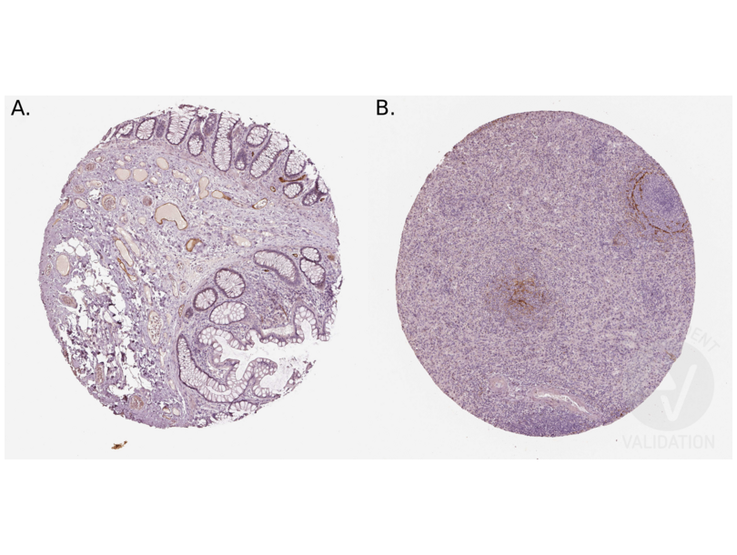
- ABIN3044076 shows nice staining in a subset of vessels (endothelial cells), present in intestine and spleen.
Validation #300031 (Immunohistochemistry)
 Validation Images
![Staining of human colon (A.) and spleen (B.) sections in a tissue microarray with ABIN3044076. See the protocol for detailed information.]() Staining of human colon (A.) and spleen (B.) sections in a tissue microarray with ABIN3044076. See the protocol for detailed information.
Protocole
Staining of human colon (A.) and spleen (B.) sections in a tissue microarray with ABIN3044076. See the protocol for detailed information.
Protocole -
- Format
- Lyophilized
- Reconstitution
- Add 0.2 mL of distilled water will yield a concentration of 500 μg/mL.
- Concentration
- 500 μg/mL
- Buffer
- Each vial contains 5 mg BSA, 0.9 mg NaCl, 0.2 mg Na2HPO4, 0.05 mg Thimerosal, 0.05 mg Sodium azide.
- Agent conservateur
- Thimerosal (Merthiolate), Sodium azide
- Précaution d'utilisation
- This product contains Sodium azide and Thimerosal (Merthiolate): POISONOUS AND HAZARDOUS SUBSTANCES which should be handled by trained staff only.
- Conseil sur la manipulation
- Avoid repeated freezing and thawing.
- Stock
- 4 °C/-20 °C
- Stockage commentaire
-
At -20°C for one year. After reconstitution, at 4°C for one month.
It can also be aliquotted and stored frozen at -20 °C for a longer time. Avoid repeated freezing and thawing. - Date de péremption
- 12 months
-
- Antigène
- MADCAM1 (Mucosal Vascular Addressin Cell Adhesion Molecule 1 (MADCAM1))
- Autre désignation
- MADCAM1 (MADCAM1 Produits)
- Synonymes
- anticorps MACAM1, anticorps AV211525, anticorps mucosal vascular addressin cell adhesion molecule 1, anticorps MADCAM1, anticorps Madcam1
- Sujet
-
MADCAM1(Mucosal Vascular Addressin Cell Adhesion Molecule 1), also known as MACAM1, is a protein that in humans is encoded by the MADCAM1 gene. By PCR-based analysis of somatic cell hybrids, Leung et al.(1997) mapped the MACAM1 gene to chromosome 19. The protein encoded by this gene is an endothelil cell adhesion molecule that interacts preferentially with the leukocyte beta7 integrin LPAM-1(alpha4 / beta7), L-selectin, and VLA-4(alpha4/beta1) on myeloid cells to direct leukocytes into mucosal and inflamed tissues. It is a member of the immunoglobulin superfamily and is similar to ICAM-1 and VCAM-1.
Synonyms: Addressin mucosal antibody|hMAdCAM 1 antibody|hMAdCAM-1 antibody|MACAM1 antibody|MADCA_HUMAN antibody|MAdCAM 1 antibody|MAdCAM-1 antibody|Madcam1 antibody|Mucosal addressin cell adhesion molecule 1 antibody|Mucosal vascular addressin cell adhesion molecule 1 antibody - UniProt
- Q13477
-


