Plakophilin 2 anticorps (AA 801-881)
 (1 reference)
 (1 validation)
-
- Antigène Voir toutes Plakophilin 2 (PKP2) Anticorps
- Plakophilin 2 (PKP2)
-
Épitope
- AA 801-881
-
Reactivité
- Humain
-
Hôte
- Lapin
-
Clonalité
- Polyclonal
-
Conjugué
- Cet anticorp Plakophilin 2 est non-conjugé
-
Application
- ELISA, Immunohistochemistry (Paraffin-embedded Sections) (IHC (p)), Immunocytochemistry (ICC), Immunohistochemistry (Frozen Sections) (IHC (fro)), Immunofluorescence (Paraffin-embedded Sections) (IF (p)), Immunofluorescence (Cultured Cells) (IF (cc))
- Réactivité croisée
- Humain
- Homologie
- Mouse,Rat,Dog,Cow,Sheep,Pig,Horse,Rabbit
- Purification
- Purified by Protein A.
- Immunogène
- KLH conjugated synthetic peptide derived from human Plakophilin 2
- Isotype
- IgG
-
anti-Plakophilin 2 (PKP2) (N-Term) antibody
PKP2 Reactivité: Humain, Rat, Souris, Boeuf (Vache), Chien, Cheval WB, IHC Hôte: Lapin Polyclonal unconjugated
anti-Plakophilin 2 (PKP2) (AA 19-183) antibodyPKP2 Reactivité: Humain WB, IHC, IF, IP Hôte: Souris Monoclonal 28-Plakophilin unconjugated
anti-Plakophilin 2 (PKP2) (AA 72-194) antibodyPKP2 Reactivité: Humain WB, IHC (p), IF Hôte: Lapin Polyclonal unconjugated
anti-Plakophilin 2 (PKP2) (C-Term) antibodyPKP2 Reactivité: Humain WB, ELISA Hôte: Lapin Polyclonal unconjugated
anti-Plakophilin 2 (PKP2) (AA 61-183) antibodyPKP2 Reactivité: Humain ELISA, IHC Hôte: Lapin Polyclonal unconjugated
anti-Plakophilin 2 (PKP2) (C-Term) antibodyPKP2 Reactivité: Humain, Rat WB, ELISA Hôte: Lapin Polyclonal unconjugated
anti-Plakophilin 2 (PKP2) (AA 838-866), (C-Term) antibodyPKP2 Reactivité: Humain WB, IHC (p) Hôte: Lapin Polyclonal RB30230 unconjugated
anti-Plakophilin 2 (PKP2) (N-Term) antibodyPKP2 Reactivité: Humain, Rat WB, ELISA, IF, ICC Hôte: Lapin Polyclonal unconjugated
anti-Plakophilin 2 (PKP2) (AA 1-350) antibodyPKP2 Reactivité: Humain, Souris WB, IHC (p), IP, FACS Hôte: Souris Monoclonal 8H6 unconjugated
anti-Plakophilin 2 (PKP2) (AA 125-139) antibodyVerified PKP2 Reactivité: Humain, Souris WB, ELISA, IHC Hôte: Chèvre Polyclonal unconjugated
-
- Indications d'application
-
ELISA 1:500-1000
IHC-P 1:200-400
IHC-F 1:100-500
IF(IHC-P) 1:50-200
IF(IHC-F) 1:50-200
IF(ICC) 1:50-200
ICC 1:100-500 - Restrictions
- For Research Use only
-
- by
- Gencardio (IDIBGI)
- No.
- #103656
- Date
- 13.03.2019
- Antigène
- PKP2
- Numéro du lot
- AD102900
- Application validée
- Immunofluorescence
- Contrôle positif
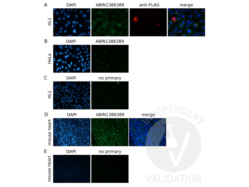
HL1 cells
HeLa cells
mouse heart tissue
- Contrôle négative
HL1 cells
HeLa cells
secondary antibody only control
- Conclusion
ABIN1386389 specifically labels the targeted antigen in mouse heart. No signal was detected in sample negative control and the secondary antibody only control.
- Anticorps primaire
- ABIN1386389
- Anticorps secondaire
- goat anti-rabbit AF488 antibody (Invitrogen, A11008, 1911238)
- Full Protocol
- HL1 and HeLa cells
- Grow HL1 cells in Claycomb medium (sigma, 51800C, SLBX8700) supplemented with 10% FBS (Gibco, 10270-106, 42G5178K), 1% Penicillin-Streptomycin (Sigma, P4333, lot 048M4874V) and 1% Glutamax (Gibco, 35050-061, lot 2037045), at 37°C and 5% CO2 in 12mm coverslips in a well of 12 well plate.
- Grow HeLa cells in MEM/EBSS medium (GE Life Science, SH30244.01, lot AC14563283) supplemented with 10% FBS (Gibco, 10270-106, lot 42G5178K), 1% Penicillin-Streptomycin (Sigma, P4333, lot 048M4874V) and 1% Glutamax (Gibco, 35050-061, lot 2037045), at 37°C and 5% CO2 in 12mm coverslips in a well of 12 well plate.
- Transfect HeLa cells with PKP2a plasmid (p915 PKP2a-pFLAG-cmv5a, provided by Dr. Green KJ) using Lipofectamine 3000 (Invitrogen, L3000-015, lot 2041107) following the manufacturer´s instructions.
- Wash cells 1x with PBS.
- Fix cells on coverslips in 4% PFA for 20min at RT.
- Wash cells 3x for 5min with PBS.
- Incubate coverslips with quench solution (PBS with 75mM glycine and 20mM NH4Cl) for 10min at RT.
- Wash cells 1x with PBS.
- Permeabilize cells and block non-specific binding with PFS-like buffer containing 0.1% BSA, 0.1% triton and 0.02% NaN3 for 30min at 37°C.
- Incubate cells with primary anti-Plakophilin 2 (PKP2) (AA 831-881) antibody (antibodies-online, ABIN1386389, lot AD102900) diluted 1:60 in PFS-like buffer or with anti-Flag-tag antibody (Sigma, monoclonal anti-flag, F316, lot SLBT7654) diluted 1:400 in PFS-like buffer ON at 4°C.
- Wash cells 3x for 5min with PBS.
- Incubate cells with secondary goat anti-rabbit AF488 antibody (Invitrogen, A11008, lot 1911238) or with secondary goat anti-mouse AF568 antibody (Invitrogen, A11031, lot 1841757) diluted 1:200 in PFS-like buffer for 30min at RT.
- Wash cells 2x for 5min with PFS-like buffer.
- Wash cells 2x for 5min with PBS.
- DAPI counterstain for 5min at RT.
- Mount coverslips on glass slides in Calbicochem (Millipore, 345789, lot 2682585).
- Image acquisition with NIS-ELEMENTS (Nikon).
- Tissue: mouse heart
- Obtain mouse heart mouse and wash it with PBS.
- Fix tissue in 15ml falcon tube with 4% PFA ON at 4°C.
- Wash the fixed heart with water.
- Embed tissue in paraffin.
- Cut the tissue using a microtrom and put it into slides.
- Desparaffinize the tissue in the slide (from xylene to water).
- Incubate the slides with retrieval solution for 30min at 95°C to permeabilize the tissue.
- Cool slides down on ice.
- Wash tissue 2x for 3min with PBS.
- Wash tissue 2x for 3min with PBS 0.1% triton.
- Block non-specific binding with PBS 0.1% triton 10% BSA for 1h at RT.
- Incubate tissue with primary anti-PKP2 (AA 831-881) antibody (antibodies-online, ABIN1386389, lot AD102900) diluted 1:100 in PBS 1% BSA solution ON at 4°C.
- Wash tissue 2x for 5min with PBS 0.1% triton.
- Incubate tissue with secondary goat anti-rabbit AF488 antibody (Invitrogen, A11008, 1911238) diluted 1:200 in PBS 1% BSA solution for 1h at RT.
- Wash cells 2x for 5min with PBS.
- Wash tissue 2x for 3min with PBS 0.1% triton.
- Wash tissue 2x for 3min with PBS.
- DAPI counterstain for 5min at RT.
- Mount slides with coverslips in Calbicochem (Millipore, 345789, lot 2682585).
- Image acquisition with NIS-ELEMENTS (Nikon).
- Notes
ABIN1386389 did not label recombinantly expressed PKP2-FLAG in transfected HL1 cells. A hypothesis could be that antibody recognizes the murine protein in mouse heart samples but not the human sequence of the vector (transfected HL1).
Validation #103656 (Immunofluorescence)
 Validation Images
![A. IF staining of cells expressing PKP2-FLAG using ABIN1386389 (green) or an anti-FLAG-tag antibody (red). Nuclear staining with DAPI (blue) (magnification 60x). B. Staining of HeLa cells not expressing PKP2-FLAG using ABIN1386389 (magnification 40x). C. Staining of cells expressing PKP2-Flag with a secondary antibody only (magnification 40x). D and E. Staining of endogenous Pkp2 in mouse heart tissue with ABIN1386389 (D) or secondary antibody only (E) (magnification 40x).]() A. IF staining of cells expressing PKP2-FLAG using ABIN1386389 (green) or an anti-FLAG-tag antibody (red). Nuclear staining with DAPI (blue) (magnification 60x). B. Staining of HeLa cells not expressing PKP2-FLAG using ABIN1386389 (magnification 40x). C. Staining of cells expressing PKP2-Flag with a secondary antibody only (magnification 40x). D and E. Staining of endogenous Pkp2 in mouse heart tissue with ABIN1386389 (D) or secondary antibody only (E) (magnification 40x).
Protocole
A. IF staining of cells expressing PKP2-FLAG using ABIN1386389 (green) or an anti-FLAG-tag antibody (red). Nuclear staining with DAPI (blue) (magnification 60x). B. Staining of HeLa cells not expressing PKP2-FLAG using ABIN1386389 (magnification 40x). C. Staining of cells expressing PKP2-Flag with a secondary antibody only (magnification 40x). D and E. Staining of endogenous Pkp2 in mouse heart tissue with ABIN1386389 (D) or secondary antibody only (E) (magnification 40x).
Protocole -
- Format
- Liquid
- Concentration
- 1 μg/μL
- Buffer
- 0.01M TBS( pH 7.4) with 1 % BSA, 0.02 % Proclin300 and 50 % Glycerol.
- Agent conservateur
- ProClin
- Précaution d'utilisation
- This product contains ProClin: a POISONOUS AND HAZARDOUS SUBSTANCE, which should be handled by trained staff only.
- Stock
- 4 °C,-20 °C
- Stockage commentaire
- Shipped at 4°C. Store at -20°C for one year. Avoid repeated freeze/thaw cycles.
- Date de péremption
- 12 months
-
-
: "Myocardial expression profiles of candidate molecules in patients with arrhythmogenic right ventricular cardiomyopathy/dysplasia compared to those with dilated cardiomyopathy and healthy controls." dans: Heart rhythm, Vol. 13, Issue 3, pp. 731-41, (2016) (PubMed).
-
: "Myocardial expression profiles of candidate molecules in patients with arrhythmogenic right ventricular cardiomyopathy/dysplasia compared to those with dilated cardiomyopathy and healthy controls." dans: Heart rhythm, Vol. 13, Issue 3, pp. 731-41, (2016) (PubMed).
-
- Antigène
- Plakophilin 2 (PKP2)
- Autre désignation
- Plakophilin 2 (PKP2 Produits)
- Synonymes
- anticorps ARVD9, anticorps 1200008D14Rik, anticorps 1200012P04Rik, anticorps AA516617, anticorps Pkp2l, anticorps plakophilin 2, anticorps PKP2, anticorps Pkp2
- Sujet
-
Synonyms: ARVD 9, ARVD-9, ARVD9, PKP 2, PKP2, PKP-2, Plakophilin-2. Plakophilin2, PKP2_HUMAN.
Background: Plakophilins 1, 2, 3 and 4 (PKP1-4) influence development and participate in linking cadherins to cytoskeletal intermediate filaments. Plakophilins 1-4 contain arm-repeat (armadillo) domains, and localize to nuclei and cell desmosomes (cell-cell junctions found in suprabasal layers of stratifying epithelia that undergo mechanical stress). Plakophilin-1 mediates increases in desmosomal protein content, desmosome assembly, and regulation of cell migration. Plakophilin-2 is important for desmosome assembly and is an essential morphogenic factor and architectural component of the heart. Plakophilin-3 plays a role in both desmosome-dependent adhesion and signaling pathways. Plakophilin-4 is a component of desmosomal adhesion plaques that regulates junctional plaque organization and cadherin function.
- ID gène
- 5318
- UniProt
- Q99959
- Pathways
- Cell-Cell Junction Organization, SARS-CoV-2 Protein Interactome, Phosphorylation & l'infection par le SRAS-CoV-2
-


