FAU anticorps (N-Term)
 (3 validations)
-
- Antigène Voir toutes FAU Anticorps
- FAU (Finkel-Biskis-Reilly Murine Sarcoma Virus (FBR-MuSV) Ubiquitously Expressed (FAU))
-
Épitope
- AA 1-30, N-Term
-
Reactivité
- Humain
-
Hôte
- Lapin
-
Clonalité
- Polyclonal
-
Conjugué
- Cet anticorp FAU est non-conjugé
-
Application
- Western Blotting (WB), Immunohistochemistry (IHC)
- Fonction
- Rabbit Anti-Human FAU (N-term) Antibody
- Immunogène
- This FAU antibody is generated from rabbits immunized with a KLH conjugated synthetic peptide between 1-30 amino acids from the N-terminal region of human FAU.
- Isotype
- Ig Fraction
-
anti-Finkel-Biskis-Reilly Murine Sarcoma Virus (FBR-MuSV) Ubiquitously Expressed (FAU) (AA 1-133) antibody
FAU Reactivité: Humain WB, ELISA, IF Hôte: Souris Monoclonal 3C10 unconjugated
anti-Finkel-Biskis-Reilly Murine Sarcoma Virus (FBR-MuSV) Ubiquitously Expressed (FAU) (C-Term) antibodyFAU Reactivité: Humain WB, IHC (p) Hôte: Lapin Polyclonal unconjugated
anti-Finkel-Biskis-Reilly Murine Sarcoma Virus (FBR-MuSV) Ubiquitously Expressed (FAU) (AA 1-59) antibodyFAU Reactivité: Humain ELISA, IHC, IF Hôte: Lapin Polyclonal unconjugated
anti-Finkel-Biskis-Reilly Murine Sarcoma Virus (FBR-MuSV) Ubiquitously Expressed (FAU) (N-Term) antibodyFAU Reactivité: Humain WB, IHC (p) Hôte: Lapin Polyclonal unconjugated
anti-Finkel-Biskis-Reilly Murine Sarcoma Virus (FBR-MuSV) Ubiquitously Expressed (FAU) (C-Term) antibodyFAU Reactivité: Humain, Souris, Rat, Lapin, Boeuf (Vache), Poisson zèbre (Danio rerio), Chien, Cobaye, Cheval, Hamster, Singe, Porc, Xenopus laevis WB, IHC, IHC (p) Hôte: Lapin Polyclonal unconjugated
anti-Finkel-Biskis-Reilly Murine Sarcoma Virus (FBR-MuSV) Ubiquitously Expressed (FAU) (AA 35-133) antibodyFAU Reactivité: Humain WB, ELISA Hôte: Souris Polyclonal unconjugated
anti-Finkel-Biskis-Reilly Murine Sarcoma Virus (FBR-MuSV) Ubiquitously Expressed (FAU) (C-Term) antibodyFAU Reactivité: Humain, Souris WB, IHC (p), EIA Hôte: Lapin Polyclonal unconjugated
anti-Finkel-Biskis-Reilly Murine Sarcoma Virus (FBR-MuSV) Ubiquitously Expressed (FAU) (AA 1-133) antibodyFAU Reactivité: Humain WB Hôte: Lapin Polyclonal unconjugated
anti-Finkel-Biskis-Reilly Murine Sarcoma Virus (FBR-MuSV) Ubiquitously Expressed (FAU) antibodyFAU Reactivité: Humain WB, FACS Hôte: Lapin Polyclonal unconjugated
anti-Finkel-Biskis-Reilly Murine Sarcoma Virus (FBR-MuSV) Ubiquitously Expressed (FAU) (AA 60-93) antibodyFAU Reactivité: Humain WB, IHC (p) Hôte: Lapin Polyclonal unconjugated
-
- Indications d'application
-
Western Blot, Immunohistochemistry
Recommended Dilutions
WB: 1:1000, IHC: 1:50-100Western blot analysis of FUBI Antibody (N-term) polyclonal antibody, which was peroxidase-conjugated to the secondary antibody, followed by DAB staining. This data demonstrates the use of this antibody for immunohistochemistry, clinical relevance has not been evaluated. - Restrictions
- For Research Use only
-
- by
- Institute for Biochemistry, Charité – Universitätsmedizin Berlin
- No.
- #103222
- Date
- 13.08.2018
- Antigène
- FAU
- Numéro du lot
- RAY8070723
- Application validée
- Western Blotting
- Contrôle positif
Mouse tissue panel
- Contrôle négative
- Conclusion
Passed, but low expression levels of endogenous FAU in most cell tissues tested.
- Anticorps primaire
- ABIN2798885
- Anticorps secondaire
- IRDye88CW goat anti-rabbit IgG (H+L) antibody (LI-COR, 926-32211)
- Full Protocol
- Remove tissues from C57BL/6 male mice of 6 to 12 months of age, anaesthetized using isofluorane and killed by dislocation of the upper cervical spinal column.
- Freeze removed tissues in liquid nitrogen.
- Homogenize brain, heart, liver, lung, kidney, spleen, skeletal muscle and fat tissue using a FastPrep instrument (Savant) in 4-6 volumes of ice-cold lysis buffer containing 20mM HEPES pH7.4, 1% Triton X-100 (v/v), 5% glycerol (v/v), 8mM EDTA, 2mM EGTA, ‘Complete’ protease inhibitor cocktail (Roche, Cat. No. 11873580001, Lot 31880700).
- Centrifuge tissue extracts at 16000rcf at for 10min at 4°C.
- Collect supernatant and determine protein concentration using Bradford reagent and bovine serum albumin as a standard.
- Denature proteins for 5min at 95°C in 1x Laemmli SDS sample buffer.
- Separate 20µg of total protein per lane on a denaturing 16% Tris-glycine mini gels (Bio-Rad, freshly cast 8.3cm x 7.3cm for Mini Protean Tetra Cell) using Tris-glycine running buffer (Tris: Fisher Scientific, BP152-5, lot 171324; Glycine: AppliChem, A1067, lot 7A015185) in a Mini Protean Tetra Cell for 75min at 160V.
- Transfer proteins onto nitrocellulose membrane membrane (LI-COR, 926-31092, lot 25452956) by semi-dry blotting (Bio-Rad Trans-Blot SD) using discontinuous Tris-CAPS buffer system.
- Block membrane either with Rotiblock (Carl Roth, A151.4, lot 297261307), 5% milk in PBS, or 1% BSA in PBS for 45min at RT.
- Incubation with primary rabbit anti-FAU antibody (antibodies-online, ABIN2798885, lot RAY8070723) diluted 1:1000 in PBS/0.1% Tween 20 ON at 4°C.
- Wash membrane 3x for 10min with PBS/0.1% Tween 20.
- Incubate membrane with secondary IRDye88CW goat anti-rabbit IgG (H+L) antibody (LI-COR, 926-32211) diluted 1:10000 in PBS/0.1% Tween 20 for 60min at RT.
- Wash membrane 3x for 10min with PBS.
- Acquire image on a LI-COR Odyssey CLX scanner.
- Notes
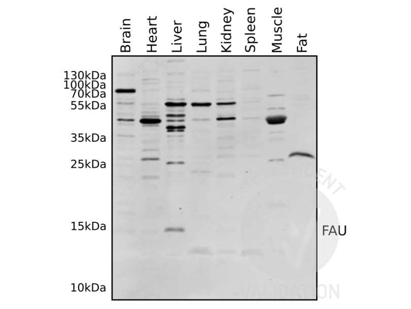
Detection of FAU (14kDa; full-length gene product: ubiquitin-like modifier FUBI + ribosomal protein S30) predominantly in liver extracts using Rotiblock as blocking reagent.
Validation #103222 (Western Blotting)
 Validation Images
![Western blot analysis of various mouse tissue lysates using ABIN2798885.]() Western blot analysis of various mouse tissue lysates using ABIN2798885.
Western blot analysis of various mouse tissue lysates using ABIN2798885.
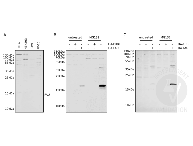
![A. Western blot analysis on cell extracts from cultured human (HeLa, HEK293), murine (RAW), and porcine (PK-15) cells using ABIN2798885. B. Western blot on extracts from HeLa cells with (+) or without (-) ectopical expression of HA-tagged FAU or FUBI. Cells were treated with proteasome inhibitor (MG132) or not (untreated). C. Western blot of the same extracts as B. using an HA-tag antibody. Blocking in all images with 5% milk in PBS.]() A. Western blot analysis on cell extracts from cultured human (HeLa, HEK293), murine (RAW), and porcine (PK-15) cells using ABIN2798885. B. Western blot on extracts from HeLa cells with (+) or without (-) ectopical expression of HA-tagged FAU or FUBI. Cells were treated with proteasome inhibitor (MG132) or not (untreated). C. Western blot of the same extracts as B. using an HA-tag antibody. Blocking in all images with 5% milk in PBS.
A. Western blot analysis on cell extracts from cultured human (HeLa, HEK293), murine (RAW), and porcine (PK-15) cells using ABIN2798885. B. Western blot on extracts from HeLa cells with (+) or without (-) ectopical expression of HA-tagged FAU or FUBI. Cells were treated with proteasome inhibitor (MG132) or not (untreated). C. Western blot of the same extracts as B. using an HA-tag antibody. Blocking in all images with 5% milk in PBS.
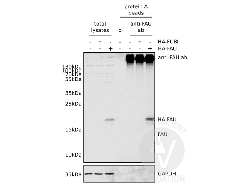
![Western blot analysis of HeLa cells expressing HA-tagged FAU (HA-FAU) or FUBI (HA-FUBI) using ABIN2798885. Extracts were submitted to immunoprecipitation using ABIN2798885 immobilized on protein A beads (protein A beads, anti-FAU ab). Total lysates or beads without immobilized antibody (protein A beads, o) were used as controls. GAPDH served as loading control.]() Western blot analysis of HeLa cells expressing HA-tagged FAU (HA-FAU) or FUBI (HA-FUBI) using ABIN2798885. Extracts were submitted to immunoprecipitation using ABIN2798885 immobilized on protein A beads (protein A beads, anti-FAU ab). Total lysates or beads without immobilized antibody (protein A beads, o) were used as controls. GAPDH served as loading control.
Protocole
Western blot analysis of HeLa cells expressing HA-tagged FAU (HA-FAU) or FUBI (HA-FUBI) using ABIN2798885. Extracts were submitted to immunoprecipitation using ABIN2798885 immobilized on protein A beads (protein A beads, anti-FAU ab). Total lysates or beads without immobilized antibody (protein A beads, o) were used as controls. GAPDH served as loading control.
Protocole - by
- Institute for Biochemistry, Charité – Universitätsmedizin Berlin
- No.
- #103412
- Date
- 13.08.2018
- Antigène
- FAU
- Numéro du lot
- RAY8070723
- Application validée
- Western Blotting
- Contrôle positif
Endogenous FAU in extracts from human (HeLa, HEK293), murine (RAW), and porcine (PK-15) secondary cells
HeLa cells transfected with HA-tagged FAU in vector pcDNA3.1
- Contrôle négative
HeLa cells transfected with empty plasmid vector pcDNA3.1
- Conclusion
Passed. ABIN2798885 specifically detects overexpressed FAU by western blotting.
- Anticorps primaire
- ABIN2798885
- Anticorps secondaire
- IRDye88CW goat anti-rabbit IgG (H+L) antibody (LI-COR, 926-32211)
- Full Protocol
- Grow HeLa, HEK293, and RAW264.7 cells in DMEM (Thermo Fisher Scientific, 41965-039, lot 1924339) supplemented with 10% FBS (Merck Millipore, S0115, lot 1248D) and antibiotics (1x Penicillin-Streptomycin, Thermo Fisher Scientific, 15140-122, lot 1970744), at 37°C and 7% CO2 in 6-well plates. Grow PK-15 cells in DMEM supplemented 5% FBS and antibiotics.
- Transfect 5x10⁵ HeLa cells/well with plasmids expressing either HA-FUBI, HA-FAU (FUBI+S30), or empty pcDNA3.1 vector using X-Treme Gene HP transfection reagent (Roche, 06366236001, lot 28049700; 3µg plasmid/well, ratio DNA to X-Treme Gene HP 1:1) following the manufacturer´s instructions. Optional: treat cells with 2.5µM proteasome inhibitor MG132 (Biozol, CAY-10012628) for 16h before lysis.
- Lyse cells in ice-cold lysis buffer containing 20mM HEPES pH 7.4, 1% Triton X-100 (v/v), 5% glycerol (v/v), 8mM EDTA, 2mM EGTA, Complete protease inhibitor cocktail (Roche, 11873580001, lot 31880700).
- Centrifuge lysates at 16000rcf for 10min at 4°C to pellet cell debris and collect supernatant.
- Determine protein concentration using Bradford reagent (Thermo Fisher Scientific, 1856210, lot MH160588) and BSA as a standard.
- Denature proteins for 5min at 95°C in 1x Laemmli SDS sample buffer.
- Separate 20µg of total protein per lane on a 16% Tris-glycine mini gels (Bio-Rad, freshly cast 8.3cm x 7.3cm for Mini Protean Tetra Cell) using Tris-glycine running buffer (Tris: Fisher Scientific, BP152-5, lot 171324; Glycine: AppliChem, A1067, lot 7A015185) in a Mini Protean Tetra Cell for 75min at 160V.
- Transfer proteins onto nitrocellulose membrane (LI-COR, 926-31092, lot 25452956) by semi-dry blotting (Bio-Rad Trans-Blot SD) using discontinuous Tris-CAPS buffer system (Bio-Rad; CAPS: Sigma-Aldrich, C2632, lot SLBR8754).
- Block membrane either with Rotiblock (Carl Roth, A151.4, lot 297261307), 5% milk in PBS, or 1% BSA in PBS for 45min at RT.
- Incubation with primary
- rabbit anti-FAU antibody (antibodies-online, ABIN2798885, lot RAY8070723) diluted 1:1000 in PBS/0.1% Tween 20 ON at 4°C or
- rabbit ant-HA antibody (Abcam, ab9110, lot GR235874-6) diluted 1:5000 in PBS/0.1% Tween 20 ON at 4°C.
- Wash membrane 3x for 10min with PBS/0.1% Tween 20.
- Incubate membrane with secondary IRDye88CW goat anti-rabbit IgG (H+L) antibody (LI-COR, 926-32211) diluted 1:10000 in PBS/0.1% Tween 20 for 1h at RT.
- Wash membrane 3x for 10min with PBS.
- Acquire image on a LI-COR Odyssey CLX scanner.
- Notes
No prominent immuno-signal for detected by Western blotting on extracts of tested secondary cell lines of human (HeLa, HEK293), murine (RAW), and porcine (PK-15) origin.
HA-tagged FAU ectopically overexpressed in HeLa cells is readily detected using anti-FAU ABIN2798885.
The presumed N-terminal cleavage product (ubiquitin-like modifier FUBI) of the FAU gene (FUBI + ribosomal protein S30) is not expressed in sufficient amounts to be detected by Western blotting. Even using a plasmid vector encoding HA-tagged ubiquitin-like modifier FUBI does not results in a proper expression of this protein.
-
- by
- Institute for Biochemistry, Charité – Universitätsmedizin Berlin
- No.
- #103413
- Date
- 13.08.2018
- Antigène
- FAU
- Numéro du lot
- RAY8070723
- Application validée
- Immunoprecipitation
- Contrôle positif
HeLa cells transfected with HA-tagged FAU in vector pcDNA3.1
- Contrôle négative
HeLa cells transfected with empty plasmid vector pcDNA3.1
- Conclusion
Passed. ABIN2798885 enriches overexpressed FAU by immunoprecipitation.
- Anticorps primaire
- ABIN2798885
- Anticorps secondaire
- IRDye88CW goat anti-rabbit IgG (H+L) antibody (LI-COR, 926-32211)
- Full Protocol
- Grow HeLa cells in DMEM (Thermo Fisher Scientific, 41965-039, lot 1924339) supplemented with 10% FBS (Merck Millipore, S0115, lot 1248D) and antibiotics (1x Penicillin-Streptomycin, Thermo Fisher Scientific, 15140-122, lot 1970744), at 37°C and 7% CO2 10cm dishes.
- Transfect 3x10⁶ cells/dish with plasmids expressing either HA-FUBI, HA-FAU (FUBI+S30), or empty pcDNA3.1 vector using X-Treme Gene HP transfection reagent (Roche, 06366236001, lot 28049700; 18µg plasmid/well, ratio DNA to X-Treme Gene HP 1:1) following the manufacturer´s instructions.
- Lyse cells in ice-cold lysis buffer containing 20mM HEPES pH 7.4, 1% Triton X-100 (v/v), 5% glycerol (v/v), 8mM EDTA, 2mM EGTA, Complete protease inhibitor cocktail (Roche, 11873580001, lot 31880700).
- Centrifuge lysates at 16000rcf for 10min at 4°C to pellet cell debris and collect supernatant.
- Determine protein concentration using Bradford reagent (Thermo Fisher Scientific, 1856210, lot MH160588) and BSA as a standard.
- Prepare aliquots of lysates for SDS-PAGE in 1x Laemmli sample buffer.
- Immobilize 10µl rabbit anti-FAU antibody (antibodies-online, ABIN2798885, RAY8070723) at 2µg/µl on 10µl SureBeads Protein A Magntic Beads (Bio-Rad, 161-4013, lot 0304JA-01) in 1ml PBS containing 0.1% Tween 20 for 30min at 4°C.
- Incubate 1mg of protein lysate in a total volume of 1 ml with 10 µl of magnetic protein A SureBeads (Bio-Rad) coupled non-covalently with anti-FAU antibody ABIN2798885 at for 1h at 4°C. Use non-coupled beads as control. Optional: use pre-immune rabbit serum as control.
- Wash beads 2x with 1ml of lysis buffer and 1x with PBS.
- Add 50µl non-reducing Laemmli buffer and incubate beads for 10min at RT to elute bound proteins.
- Separate 20µg of total protein and 1/5 of the IP sample per lane on a 16% Tris-glycine mini gels (Bio-Rad, freshly cast 8.3cm x 7.3cm for Mini Protean Tetra Cell) using Tris-glycine running buffer (Tris: Fisher Scientific, BP152-5, lot 171324; Glycine: AppliChem, A1067, lot 7A015185) in a Mini Protean Tetra Cell for 75min at 160 V.
- Transfer proteins onto nitrocellulose membrane (LI-COR, 926-31092, lot 25452956) by semi-dry blotting (Bio-Rad Trans-Blot SD) using discontinuous Tris-CAPS buffer system (Bio-Rad; CAPS: Sigma-Aldrich, C2632, lot SLBR8754).
- Block membrane with 5% milk in PBS for 45min at RT.
- Incubation with primary
- rabbit anti-FAU antibody ABIN2798885 diluted 1:1000 in PBS/0.1% Tween 20 ON at 4°C or
- rabbit anti-GAPDH antibody (Santa Cruz, sc-25778).
- Wash membrane 3x for 10min with PBS/0.1% Tween 20.
- Incubate membrane with secondary IRDye88CW goat anti-rabbit IgG (H+L) antibody (LI-COR, 926-32211) diluted 1:10000 in PBS/0.1% Tween 20 for 1h at RT.
- Wash membrane 3x for 10 min with PBS.
- Acquire image on a LI-COR Odyssey CLX scanner.
- Notes
ABIN2798885 readily immunoprecipitates FAU protein, although overexpression might be required. Expression levels of endogenous FAU in HeLa cells appear to be too low to achieve proper enrichment by immunoprecipitation.
The presumed N-terminal cleavage product (ubiquitin-like modifier FUBI) of the FAU gene (FUBI + ribosomal protein S30) is not expressed in sufficient amounts to be detected by Western blotting. Even using a plasmid vector encoding HA-tagged ubiquitin-like modifier FUBI does not results in a proper expression of this protein.
Validation #103413 (Immunoprecipitation)
 Validation Images
![Western blot analysis of various mouse tissue lysates using ABIN2798885.]() Western blot analysis of various mouse tissue lysates using ABIN2798885.
Western blot analysis of various mouse tissue lysates using ABIN2798885.
![A. Western blot analysis on cell extracts from cultured human (HeLa, HEK293), murine (RAW), and porcine (PK-15) cells using ABIN2798885. B. Western blot on extracts from HeLa cells with (+) or without (-) ectopical expression of HA-tagged FAU or FUBI. Cells were treated with proteasome inhibitor (MG132) or not (untreated). C. Western blot of the same extracts as B. using an HA-tag antibody. Blocking in all images with 5% milk in PBS.]() A. Western blot analysis on cell extracts from cultured human (HeLa, HEK293), murine (RAW), and porcine (PK-15) cells using ABIN2798885. B. Western blot on extracts from HeLa cells with (+) or without (-) ectopical expression of HA-tagged FAU or FUBI. Cells were treated with proteasome inhibitor (MG132) or not (untreated). C. Western blot of the same extracts as B. using an HA-tag antibody. Blocking in all images with 5% milk in PBS.
A. Western blot analysis on cell extracts from cultured human (HeLa, HEK293), murine (RAW), and porcine (PK-15) cells using ABIN2798885. B. Western blot on extracts from HeLa cells with (+) or without (-) ectopical expression of HA-tagged FAU or FUBI. Cells were treated with proteasome inhibitor (MG132) or not (untreated). C. Western blot of the same extracts as B. using an HA-tag antibody. Blocking in all images with 5% milk in PBS.
![Western blot analysis of HeLa cells expressing HA-tagged FAU (HA-FAU) or FUBI (HA-FUBI) using ABIN2798885. Extracts were submitted to immunoprecipitation using ABIN2798885 immobilized on protein A beads (protein A beads, anti-FAU ab). Total lysates or beads without immobilized antibody (protein A beads, o) were used as controls. GAPDH served as loading control.]() Western blot analysis of HeLa cells expressing HA-tagged FAU (HA-FAU) or FUBI (HA-FUBI) using ABIN2798885. Extracts were submitted to immunoprecipitation using ABIN2798885 immobilized on protein A beads (protein A beads, anti-FAU ab). Total lysates or beads without immobilized antibody (protein A beads, o) were used as controls. GAPDH served as loading control.
Protocole
Western blot analysis of HeLa cells expressing HA-tagged FAU (HA-FAU) or FUBI (HA-FUBI) using ABIN2798885. Extracts were submitted to immunoprecipitation using ABIN2798885 immobilized on protein A beads (protein A beads, anti-FAU ab). Total lysates or beads without immobilized antibody (protein A beads, o) were used as controls. GAPDH served as loading control.
Protocole -
- Format
- Liquid
- Concentration
- 2.0 mg/mL
- Stock
- 4 °C,-20 °C
- Stockage commentaire
- 2-8°C (short-term), -20°C (long-term)
-
- Antigène
- FAU (Finkel-Biskis-Reilly Murine Sarcoma Virus (FBR-MuSV) Ubiquitously Expressed (FAU))
- Autre désignation
- FAU (FAU Produits)
- Synonymes
- anticorps FAU1, anticorps Fub1, anticorps Fubi, anticorps MNSFbeta, anticorps RPS30, anticorps S30, anticorps asr1, anticorps Asr1, anticorps MGC73171, anticorps zgc:73171, anticorps FAU, ubiquitin like and ribosomal protein S30 fusion, anticorps Finkel-Biskis-Reilly murine sarcoma virus (FBR-MuSV) ubiquitously expressed (fox derived), anticorps Finkel-Biskis-Reilly murine sarcoma virus (FBR-MuSV) ubiquitously expressed a, anticorps FAU, anticorps Fau, anticorps faua
- Sujet
-
Target Description: FUBI is the cellular homolog of the fox sequence in the Finkel-Biskis-Reilly murine sarcoma virus (FBR-MuSV). It is a fusion protein consisting of the ubiquitin-like protein fubi at the N terminus and ribosomal protein S30 at the C terminus. It has been proposed that the fusion protein is post-translationally processed to generate free fubi and free ribosomal protein S30. Fubi is a member of the ubiquitin family, and ribosomal protein S30 belongs to the S30E family of ribosomal proteins. Whereas the function of fubi is currently unknown, ribosomal protein S30 is a component of the 40S subunit of the cytoplasmic ribosome.
Gene Symbol: FAU
- Poids moléculaire
- 7760 Da
- ID gène
- 2197
- UniProt
- P35544
-
 Western blot analysis of various mouse tissue lysates using ABIN2798885.
 A. Western blot analysis on cell extracts from cultured human (HeLa, HEK293), murine (RAW), and porcine (PK-15) cells using ABIN2798885. B. Western blot on extracts from HeLa cells with (+) or without (-) ectopical expression of HA-tagged FAU or FUBI. Cells were treated with proteasome inhibitor (MG132) or not (untreated). C. Western blot of the same extracts as B. using an HA-tag antibody. Blocking in all images with 5% milk in PBS.
 Western blot analysis of HeLa cells expressing HA-tagged FAU (HA-FAU) or FUBI (HA-FUBI) using ABIN2798885. Extracts were submitted to immunoprecipitation using ABIN2798885 immobilized on protein A beads (protein A beads, anti-FAU ab). Total lysates or beads without immobilized antibody (protein A beads, o) were used as controls. GAPDH served as loading control.


