CTNNB1 anticorps (N-Term)
 (8 références)
 (1 validation)
-
- Highlights
-
- L'anticorps de lapin contre la bêta-caténine détecte de manière fiable les cibles de la bêta-caténine dans la signalisation Wnt en utilisant CUT&RUN, ICC, IF, WB, ELISA.
- Convient parfaitement à l'imagerie des jonctions d'adhérence en IHC/IF.
- Détection hautement spécifique de la bêta-caténine en WB.
- Antigène Voir toutes CTNNB1 Anticorps
- CTNNB1 (Catenin (Cadherin-Associated Protein), beta 1, 88kDa (CTNNB1))
-
Épitope
- N-Term
-
Reactivité
- Humain
-
Hôte
- Lapin
-
Clonalité
- Polyclonal
-
Conjugué
- Cet anticorp CTNNB1 est non-conjugé
-
Application
- Western Blotting (WB), Immunohistochemistry (IHC), Immunofluorescence (IF), Immunohistochemistry (Paraffin-embedded Sections) (IHC (p)), Flow Cytometry (FACS), Immunoprecipitation (IP), Immunocytochemistry (ICC), Immunohistochemistry (Frozen Sections) (IHC (fro)), Chromatin Immunoprecipitation (ChIP), Immunohistochemistry (Whole Mount) (IHC (wm)), Cleavage Under Targets and Release Using Nuclease (CUT&RUN)
- Réactivité croisée
- Cat, Chien, Humain, Souris, Lapin, Rat, Poisson zèbre (Danio rerio)
- Attributs du produit
-
Rabbit Polyclonal antibody to beta Catenin (catenin (cadherin-associated protein), beta 1, 88 kDa)
beta Catenin antibody [N1N2-2], N-term - Purification
- Purified by antigen-affinity chromatography.
- Classe de qualité
- KO Validated
- Immunogène
- Recombinant protein encompassing a sequence within the N-terminus region of human beta Catenin. The exact sequence is proprietary.
- Isotype
- IgG
- Informations sur le produit
-
À quoi peut servir l'anticorps ABIN2855042 contre la bêta-caténine ? Cet anticorps polyclonal de lapin anti-bêta caténine non conjugué convient à la détection de la bêta caténine (CTNNB1, Caténine bêta-1, β-Catenin) par immunoprécipitation de la chromatine (ChIP), CUT&RUN, immunofluorescence, ELISA, IHC et Western blotting. Il a été validé pour les espèces humaine, de souris et de rat. Les réactivités supplémentaires prédites sont celles du poulet, du porc, du Xenopus laevis, du singe rhésus et du poisson zèbre.
Quelles sont les données de validation disponibles pour cet anticorps anti-caténine bêta ? L'anticorps beta Catenin a actuellement une validation indépendante pour CUT&RUN (Cleavage Under Targets and Release Using Nuclease) qui peut être lue avec le protocole ci-dessous. Il a été utilisé dans sept publications, qui sont également indiquées et peuvent être trouvées sur PubMed. L'anticorps beta Catenin / β-Catenin de lapin a 16 images de produit démontrant sa performance dans CUT&RUN, Western blot, ICC / IF et IHC. Utilisez notre anticorps de haute qualité contre la bêta caténine pour détecter de manière fiable la bêta caténine pour l'homme et plusieurs autres espèces.
Quelle est la fonction de la bêta-caténine ? La bêta caténine est un composant clé en aval de la voie de signalisation canonique Wnt. En l'absence de Wnt, forme un complexe avec AXIN1, AXIN2, APC, CSNK1A1 et GSK3B qui favorise la phosphorylation sur les résidus Ser et Thr N-terminaux et l'ubiquitination de la bêta Caténine via BTRC et sa dégradation ultérieure par le protéasome. En présence du ligand Wnt, la bêta caténine n'est pas ubiquitinée et s'accumule dans le noyau, où elle agit comme un coactivateur pour les facteurs de transcription de la famille TCF/LEF, conduisant à l'activation des gènes sensibles à Wnt. (UniProt) C'est une cible difficile pour ChIP-seq, mais vous pouvez utiliser notre anticorps beta Catenin dans CUT&RUN.
-
anti-Catenin (Cadherin-Associated Protein), beta 1, 88kDa (CTNNB1) (AA 692-721), (C-Term) antibody
CTNNB1 Reactivité: Humain WB, IF, IHC (p), FACS Hôte: Lapin Polyclonal RB41124 unconjugated
anti-Catenin (Cadherin-Associated Protein), beta 1, 88kDa (CTNNB1) (N-Term) antibodyCTNNB1 Reactivité: Humain, Souris, Rat WB, IHC, ELISA, IF, ICC Hôte: Lapin Polyclonal unconjugated
anti-Catenin (Cadherin-Associated Protein), beta 1, 88kDa (CTNNB1) (AA 2-781) antibodyCTNNB1 Reactivité: Humain WB, IHC, ELISA, IF, IP Hôte: Lapin Polyclonal unconjugated
anti-Catenin (Cadherin-Associated Protein), beta 1, 88kDa (CTNNB1) (AA 78-106), (N-Term) antibodyCTNNB1 Reactivité: Humain WB, IF, IHC (p), FACS Hôte: Lapin Polyclonal RB41155 unconjugated
anti-Catenin (Cadherin-Associated Protein), beta 1, 88kDa (CTNNB1) (AA 1-100) antibodyCTNNB1 Reactivité: Humain, Souris WB, IHC, ELISA, FACS, ICC, Neut Hôte: Souris Monoclonal 7C5A2 unconjugated
anti-Catenin (Cadherin-Associated Protein), beta 1, 88kDa (CTNNB1) (AA 632-781) antibodyCTNNB1 Reactivité: Humain, Souris, Rat, Singe WB, IHC, ELISA, FACS Hôte: Souris Monoclonal 5B6E9 unconjugated
anti-Catenin (Cadherin-Associated Protein), beta 1, 88kDa (CTNNB1) antibodyCTNNB1 Reactivité: Humain WB, IHC, IF, FACS, StM Hôte: Lapin Monoclonal CTNNB1-2030R unconjugated Recombinant Antibody
anti-Catenin (Cadherin-Associated Protein), beta 1, 88kDa (CTNNB1) (AA 682-781) antibodyCTNNB1 Reactivité: Humain WB, ELISA, IF, PLA, RNAi Hôte: Souris Monoclonal 1C9 unconjugated
anti-Catenin (Cadherin-Associated Protein), beta 1, 88kDa (CTNNB1) antibodyCTNNB1 Reactivité: Humain, Chien WB, IHC, IF, IHC (p), FACS Hôte: Souris Monoclonal 1E10 unconjugated
anti-Catenin (Cadherin-Associated Protein), beta 1, 88kDa (CTNNB1) antibodyCTNNB1 Reactivité: Humain, Rat, Singe WB, IHC, IF, IHC (p), FACS Hôte: Souris Monoclonal 12H7 unconjugated
-
- Indications d'application
- WB: 1:500-1:20000. ICC/IF: 1:100-1:1000. IHC-P: 1:100-1:1000. FACS: 1:50-1:200. IP: 1:50-1:100. Optimal dilutions/concentrations should be determined by the researcher. Not tested in other applications.
- Commentaires
-
Positive Control: Mouse brain , PC-12 , HeLa
Validation: Comparison, KO/KD, Orthogonal
- Restrictions
- For Research Use only
-
- by
- Cantù Lab, Gene Regulation during Development and Disease, Linköping University
- No.
- #104235
- Date
- 28.06.2021
- Antigène
- beta Catenin
- Numéro du lot
- 42536
- Application validée
- Cleavage Under Targets and Release Using Nuclease
- Contrôle positif
Recombinant anti-H3K27me3 CUT&RUN Positive Control antibody (antibodies-online, ABIN6923144)
- Contrôle négative
- Monoclonal anti-FLAG (Sigma-Aldrich, F3165)
- Conclusion
Passed. ABIN2855042 allows for beta Catenin-targeted digestion using CUT&RUN on HEK-293 nuclei.
- Anticorps primaire
- ABIN2855042
- Anticorps secondaire
- Full Protocol
- Cell harvest
- Harvest 500,000 HEK293T cells per antibody to be used at RT.
- Centrifuge cell solution 3 min at 600 x g at RT.
- Remove the liquid carefully.
- Gently resuspend cells in 1 mL Wash Buffer (20 mM HEPES pH 7.5, 150 mM NaCl, 0.5 mM Spermidine, Roche Complete Protease Inhibitor EDTA-free) by pipetting and transfer cell solution to a 2 mL microcentrifuge tube.
- Centrifuge cell solution 3 min at 600 x g at RT and discard the supernatant.
- Repeat twice for a total of three washes.
- Resuspend cell pellet in 1 mL Wash Buffer by gently pipetting.
- Concanavalin A beads preparation
- Prepare one 1.5 mL microcentrifuge tube.
- Gently resuspend the magnetic Concanavalin A Beads (antibodies-online, ABIN6923139).
- Pipette 40 µL Con A Beads slurry for each sample into the 1.5 mL microcentrifuge tube.
- Place the tube on a magnet stand until the fluid is clear. Remove the liquid carefully.
- Remove the microcentrifuge tube from the magnetic stand.
- Pipette 1 mL Binding Buffer (20 mM HEPES pH 7.5, 10 mM KCl, 1 mM CaCl2, 1 mM MnCl2) into each tube and resuspend ConA beads by gentle pipetting.
- Spin down the liquid from the lid with a quick pulse in a table-top centrifuge.
- Place the tubes on a magnet stand until the fluid is clear. Remove the liquid carefully.
- Remove the microcentrifuge tube from the magnetic stand.
- Repeat twice for a total of three washes.
- Gently resuspend the ConA Beads in a volume of Binding Buffer corresponding to the original volume of bead slurry, i.e. 40 µL per sample.
- Nuclei immobilization – binding to Concanavalin A beads
- Carefully vortex the cell suspension and add 10 µL of the Con A beads in Binding Buffer to the cell suspension for each sample.
- Close tube tightly and rotate for 10 min at RT.
- Primary antibody binding
- oDivide nuclei suspension into separate 2 mL microcentrifuge tubes, one for each antibody (500,000 nuclei per sample).
- Place the microcentrifuge tubes on a magnetic stand until the fluid is clear. Remove the liquid carefully.
- Remove the microcentrifuge tubes from the magnetic stand.
- Place each tube at a low angle on the vortex mixer set to a low speed and add 150 µL Digitonin Wash buffer (wash buffer with 0.025% (wt/vol) Digitonin) supplemented with 2 mM EDTA.
- Gently vortex the microcentrifuge tubes until the beads are resuspended.
- Add 1.5 µL antibody (anti-beta Catenin antibody ABIN2855042, anti-H3K27me3 positive control antibody ABIN6923144, and anti-FLAG tag antibody negative control) to the respective tube, corresponding to a 1:100 dilution.
- Rotate the microcentrifuge tubes ON at 4 °C.
- Spin down the liquid and place the tubes on a magnet stand until the fluid is clear. Remove the liquid carefully.
- Remove the microcentrifuge tubes from the magnetic stand.
- Resuspend with 1 mL Digitonin Wash Buffer and mix by inversion. If clumping occurs, gently remove the clumps with a 1 ml pipette tip.
- Repeat once for a total of two washes.
- pAG-MNase Binding
- Place the tubes on a magnet stand until the fluid is clear. Remove the liquid carefully.
- Remove the microcentrifuge tubes from the magnetic stand.
- o Vortex the sample at low speed and add 2.5 µL (0.05 volumes) CUTANA pAG-MNase for ChIC/CUT&RUN Assays (ABIN6950951) in 50 µL Digitonin Wash Buffer per sample, gently resuspending the beads by pipetting.
- Rotate the microcentrifuge tubes for 1 h at 4 °C.
- Spin down the liquid and place the tubes on a magnet stand until the fluid is clear. Remove the liquid carefully.
- Remove the microcentrifuge tubes from the magnetic stand.
- Resuspend with 1 mL Digitonin Wash Buffer and mix by inversion. If clumping occurs, gently remove the clumps with a 1 mL pipette tip.
- Repeat once for a total of two washes.
- MNase digestion and release of pAG-MNase-antibody-chromatin complexes
- Spin down the liquid from the lid with a quick pulse in a table-top centrifuge.
- Place the tubes on a magnet stand until the fluid is clear. Remove the liquid carefully.
- Place each tube at a low angle on the vortex mixer set to a low speed and add 100 μL Digitonin Wash buffer per sample along the side of the tube.
- Place tubes in a heat block, kept on ice, and allow to chill.
- Add 2 μL 0.1 M CaCl2 to each sample.
- Incubate tubes at 0 °C for 30 min.
- Add 100 μL 2xSTOP buffer (340 mM NaCl, 20 mM EDTA, 4 mM EGTA, 0.05% (wt/vol) Digitonin, 100 μg/mL RNAse A, 50 μg/mL Glycogen).
- Incubate tubes at 37 °C for 30 min.
- Place the tubes on a magnet stand until the fluid is clear.
- Transfer the supernatant containing the pA-MNase-bound digested chromatin fragments to fresh 1.5 mL microcentrifuge tubes.
- DNA extraction
- Add 2 µL 10% SDS to a final concentration of 0.1% and 2.5 µL Proteinase K (20 mg/mL) to each supernatant.
- Gently vortex tubes at a low speed of approximately 1,100 rpm.
- Incubate tubes at 50 °C for 1 h.
- Add 200 µL PCI to tube.
- Vortex tubes thoroughly at high speed until the liquid appears milky.
- Centrifuge tubes in a tabletop centrifuge at 16,000 x g at RT for 5 min.
- Carefully transfer to upper aqueous phase to a fresh 1.5 mL microcentrifuge tube containing 2 µL glycogen (diluted 1:10 to 2 mg/mL from the 20 mg/mL stock solution).
- Add 20 µL 3 M NaOAc pH 5.2.
- Add 400 µL 100% ethanol.
- Place tubes for at -20 °C ON.
- Centrifuge tubes in a tabletop centrifuge at 16,000 x g at 4 °C for 5min.
- Remove the liquid carefully with a pipette.
- Wash pellet with 1ml 70% ethanol.
- Centrifuge tubes in a tabletop centrifuge at 16,000 x g at 4 °C for 1 min.
- Remove the liquid carefully with a pipette.
- Air-dry the pellet, then dissolve in 30 µL 1 mM Tris-HCl, 0.1 mM EDTA.
- Library preparation and sequencing
- Prepare libraries using KAPA HyperPrep Kit using KAPA Dual-Indexed adapters according to protocol.
- Sequence libraries on an Illumina NextSeq 500 sequencer, using a NextSeq 500/550 High Output Kit v2.5 (75 Cycles), 36bp PE.
- Peak calling
- Map reads to the GRCm38 (mm10) mouse genome using Bowtie2 with options: --local --very-sensitive- local --no-unal --no-mixed --no-discordant.
- Call peaks using MACS2 with options -f BAMPE --keep-dup all –nomodel.
- Notes
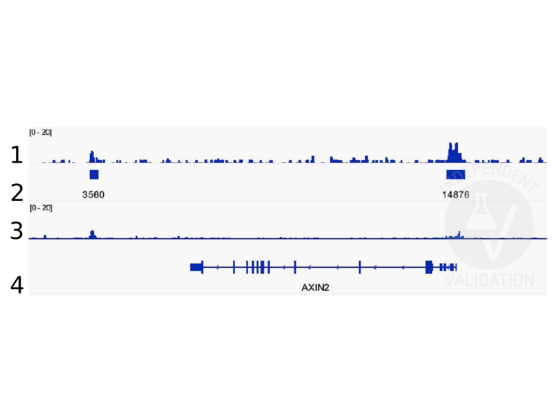
Peaks generated using ABIN2855074 for CUT&RUN aligned well with beta Catenin ChIP-seq tracks(Doumpas et al., 2019).
Validation #104235 (Cleavage Under Targets and Release Using Nuclease)
 Validation Images
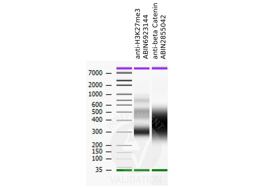
![Bioanalyzer profiles comparing fragment size distributions between reads obtained from CUT&RUN using an anti-H3K27me3 CUT&RUN Positive Control antibody ABIN6923144 and anti-beta Catenin ABIN2855042 after library preparation.]() Bioanalyzer profiles comparing fragment size distributions between reads obtained from CUT&RUN using an anti-H3K27me3 CUT&RUN Positive Control antibody ABIN6923144 and anti-beta Catenin ABIN2855042 after library preparation.
Bioanalyzer profiles comparing fragment size distributions between reads obtained from CUT&RUN using an anti-H3K27me3 CUT&RUN Positive Control antibody ABIN6923144 and anti-beta Catenin ABIN2855042 after library preparation.
![Alignment tracks from CUT&RUN targeting beta Catenin in HEK293T cells. 1. Alignment track for CUT&RUN reads obtained using anti-beta Catenin antibody ABIN2855042 in HEK293T cells. 2. Peaks called by SEACR from CUT&RUN data using anti-beta Catenin antibody ABIN2855042. 3. Peak track from ChIP-seq targeting beta Catenin in HEK293T cells (Doumpas et al., 2019). 4. Refseq track showing the AXIN2 gene on (chr17:65528563-65561648).]() Alignment tracks from CUT&RUN targeting beta Catenin in HEK293T cells. 1. Alignment track for CUT&RUN reads obtained using anti-beta Catenin antibody ABIN2855042 in HEK293T cells. 2. Peaks called by SEACR from CUT&RUN data using anti-beta Catenin antibody ABIN2855042. 3. Peak track from ChIP-seq targeting beta Catenin in HEK293T cells (Doumpas et al., 2019). 4. Refseq track showing the AXIN2 gene on (chr17:65528563-65561648).
Protocole
Alignment tracks from CUT&RUN targeting beta Catenin in HEK293T cells. 1. Alignment track for CUT&RUN reads obtained using anti-beta Catenin antibody ABIN2855042 in HEK293T cells. 2. Peaks called by SEACR from CUT&RUN data using anti-beta Catenin antibody ABIN2855042. 3. Peak track from ChIP-seq targeting beta Catenin in HEK293T cells (Doumpas et al., 2019). 4. Refseq track showing the AXIN2 gene on (chr17:65528563-65561648).
Protocole -
- Format
- Liquid
- Concentration
- 0.07 mg/mL
- Buffer
- 1XPBS ( pH 7), 1 % BSA, 20 % Glycerol, 0.025 % ProClin 300
- Agent conservateur
- ProClin
- Précaution d'utilisation
- This product contains ProClin: a POISONOUS AND HAZARDOUS SUBSTANCE which should be handled by trained staff only.
- Stock
- 4 °C,-20 °C
- Stockage commentaire
- Store as concentrated solution. Centrifuge briefly prior to opening vial. For short-term storage (1-2 weeks), store at 4°C. For long-term storage, aliquot and store at -20°C or below. Avoid multiple freeze-thaw cycles.
-
-
: "Acute Intoxication With Alcohol Reduces Trauma-Induced Proinflammatory Response and Barrier Breakdown in the Lung via the Wnt/β-Catenin Signaling Pathway." dans: Frontiers in immunology, Vol. 13, pp. 866925, (2022) (PubMed).
: "A new cut&run low volume-urea (LoV-U) protocol optimized for transcriptional co-factors uncovers Wnt/b-catenin tissue-specific genomic targets." dans: Development (Cambridge, England), (2022) (PubMed).
: "Identification of candidate lncRNAs and circRNAs regulating WNT3/β-catenin signaling in essential hypertension." dans: Aging, Vol. 12, Issue 9, pp. 8261-8288, (2020) (PubMed).
: "A novel c-Kit/phospho-prohibitin axis enhances ovarian cancer stemness and chemoresistance via Notch3-PBX1 and β-catenin-ABCG2 signaling." dans: Journal of biomedical science, Vol. 27, Issue 1, pp. 42, (2020) (PubMed).
: "Small G protein signalling modulator 2 (SGSM2) is involved in oestrogen receptor-positive breast cancer metastasis through enhancement of migratory cell adhesion via interaction with E-cadherin." dans: Cell adhesion & migration, Vol. 13, Issue 1, pp. 120-137, (2019) (PubMed).
: "Targeting the Wnt/β-catenin pathway in human osteosarcoma cells." dans: Oncotarget, Vol. 9, Issue 95, pp. 36780-36792, (2018) (PubMed).
: "ST6GALNAC1 plays important roles in enhancing cancer stem phenotypes of colorectal cancer via the Akt pathway." dans: Oncotarget, Vol. 8, Issue 68, pp. 112550-112564, (2017) (PubMed).
: "Control of the negative IRES trans-acting factor KHSRP by ubiquitination." dans: Nucleic acids research, Vol. 45, Issue 1, pp. 271-287, (2017) (PubMed).
-
: "Acute Intoxication With Alcohol Reduces Trauma-Induced Proinflammatory Response and Barrier Breakdown in the Lung via the Wnt/β-Catenin Signaling Pathway." dans: Frontiers in immunology, Vol. 13, pp. 866925, (2022) (PubMed).
-
- Antigène
- CTNNB1 (Catenin (Cadherin-Associated Protein), beta 1, 88kDa (CTNNB1))
- Autre désignation
- catenin beta 1 (CTNNB1 Produits)
- Synonymes
- anticorps Jup, anticorps LOC100217813, anticorps CTNNB1, anticorps CTNNB, anticorps Bfc, anticorps Catnb, anticorps Mesc, anticorps ctnnb, anticorps id:ibd2058, anticorps wu:fb73e10, anticorps wu:fi81c06, anticorps wu:fk25h01, anticorps ctnnb1, anticorps CHBCAT, anticorps MRD19, anticorps armadillo, anticorps beta-catenin, anticorps catenin beta 1, anticorps catenin (cadherin associated protein), beta 1, anticorps catenin (cadherin-associated protein), beta 1, anticorps catenin beta 1 S homeolog, anticorps catenin beta 1 L homeolog, anticorps CTNNB1, anticorps Ctnnb1, anticorps ctnnb1, anticorps ctnnb1.S, anticorps ctnnb1.L
- Sujet
-
Beta-catenin is an adherens junction protein. Adherens junctions (AJs, also called the zonula adherens) are critical for the establishment and maintenance of epithelial layers, such as those lining organ surfaces. AJs mediate adhesion between cells, communicate a signal that neighboring cells are present, and anchor the actin cytoskeleton. In serving these roles, AJs regulate normal cell growth and behavior. At several stages of embryogenesis, wound healing, and tumor cell metastasis, cells form and leave epithelia. This process, which involves the disruption and reestablishment of epithelial cell-cell contacts, may be regulated by the disassembly and assembly of AJs. AJs may also function in the transmission of the 'contact inhibition' signal, which instructs cells to stop dividing once an epithelial sheet is complete.[supplied by OMIM]
Cellular Localization: Cytoplasm , Nucleus , cytoskeleton - Poids moléculaire
- 85 kDa
- ID gène
- 1499
- UniProt
- P35222
- Pathways
- Signalisation WNT, Intracellular Steroid Hormone Receptor Signaling Pathway, Peptide Hormone Metabolism, Regulation of Muscle Cell Differentiation, Cell-Cell Junction Organization, Tube Formation, Maintenance of Protein Location, Signaling Events mediated by VEGFR1 and VEGFR2
-


