Androgen Receptor anticorps
 (1 validation)
-
- Antigène Voir toutes Androgen Receptor (AR) Anticorps
- Androgen Receptor (AR)
-
Reactivité
- Humain, Souris, Rat, Singe
-
Hôte
- Lapin
-
Clonalité
- Polyclonal
-
Conjugué
- Cet anticorp Androgen Receptor est non-conjugé
-
Application
- Western Blotting (WB), Immunohistochemistry (IHC), Immunofluorescence (IF), Immunochromatography (IC)
- Specificité
- Recognizes endogenous levels of Androgen Receptor protein.
- Attributs du produit
- Rabbit polyclonal antibody to Androgen Receptor
- Purification
- The antibody was purified by immunogen affinity chromatography.
- Immunogène
- KLH-conjugated synthetic peptide encompassing a sequence within the center region of human Androgen Receptor.
- Top Product
- Discover our top product AR Anticorps primaire
-
anti-Androgen Receptor (AR) antibody
AR Reactivité: Humain, Souris, Rat WB, IHC, IF, ICC Hôte: Lapin Polyclonal unconjugated
anti-Androgen Receptor (AR) (N-Term) antibodyAR Reactivité: Humain WB, IF, IHC (p), ICC Hôte: Lapin Polyclonal unconjugated
anti-Androgen Receptor (AR) antibodyAR Reactivité: Humain WB, ELISA, IHC Hôte: Souris Monoclonal 2H8 unconjugated
anti-Androgen Receptor (AR) (AA 491-679) antibodyAR Reactivité: Rat WB, IHC, IP, ICC Hôte: Souris Monoclonal D4 unconjugated
anti-Androgen Receptor (AR) (AA 221-320) antibodyAR Reactivité: Humain WB, ELISA, IHC (p), PLA Hôte: Souris Monoclonal 1G3 unconjugated
anti-Androgen Receptor (AR) (N-Term) antibodyVerified AR Reactivité: Humain WB, ELISA, IHC, IF, ChIP Hôte: Chèvre Polyclonal unconjugated
anti-Androgen Receptor (AR) (AA 15-246) antibodyAR Reactivité: Humain WB, IHC, IP, ICC Hôte: Lapin Polyclonal unconjugated
anti-Androgen Receptor (AR) (AA 365-392) antibodyAR Reactivité: Humain, Souris WB, ELISA, IHC Hôte: Lapin Polyclonal unconjugated
anti-Androgen Receptor (AR) (pSer578) antibodyAR Reactivité: Humain, Souris, Rat IHC (p), IF (p) Hôte: Lapin Polyclonal unconjugated
anti-Androgen Receptor (AR) (AA 825-919) antibodyAR Reactivité: Humain, Souris, Rat, Porc, Chévre WB, ELISA, IHC (p), IF (p), IF (cc), IHC (fro) Hôte: Lapin Polyclonal unconjugated
-
- Indications d'application
- WB (1:500 - 1:1000), IH (1:100 - 1:200), IF/IC (1:100 - 1:500)
- Restrictions
- For Research Use only
-
- by
- School of Science and Engineering, Tulane University
- No.
- #101650
- Date
- 21.12.2017
- Antigène
- Androgen Receptor
- Numéro du lot
- CP1D10A
- Application validée
- Immunofluorescence
- Contrôle positif
- prostate from inducible ARlacZ mouse
- Contrôle négative
- secondary antibody only
- Conclusion
Passed. Despite some weak non-specific staining, this antibody robustly labels the AR at a dilution of 1:100.
- Anticorps primaire
- ABIN2957859
- Anticorps secondaire
- goat anti-rabbit Cy3 conjugated antibody (Jackson ImmunoResearch)
- Full Protocol
- Prostate Tissue from ARlacz transgenic mouse or wild type mouse is fixed via transcardial perfusion with 10% formalin and postfixed at 4°C overnight. Tissue from the transgenic mouse is used for a positive control while tissue from the wild-type mouse is used with the AR antibody.
- Dewaxing and rehydration:
- CitriSolv (Decon Laboratories, 1601H, 11068) 2x for 5min.
- 100% ethanol 2x for 3min.
- 95% ethanol 2x for 3min.
- 70% ethanol 2x for 3min.
- Rinse well in distilled H2O.
- HIER:
- Dilute 10x citrate buffer pH6.0 1:10 with dH2O and place in cases.
- Place one slide into each case in the preheated pressure cooker (Hamilton Beach).
- Antigen unmasking for 40min at 92-99°C.
- Remove slides from pressure cooker (but leave them in the cases with citrate buffer). Let slides cool down for 20min to approximately 30°C.
- Wash sections 2x for 5min with PBST rocking in a coplin jar.
- Wash sections for 5min with PBS rocking in a coplin jar.
- Permeabilize sections 10min in PBS + 0.2 Triton X-100 rocking in a coplin jar.
- Wash sections 3x for 5min with PBST rocking in a coplin jar.
- Aspirate liquid, circle tissue with a PAP pen and let dry.
- Block each section with 200µl blocking solution (PBS containing 1% BSA, 5% normal goat serum, and 0.02% Triton X-100) for 1h at RT.
- Aspirate blocking solution.
- Incubate sections with 100µl primary
- rabbit anti-androgen receptor antibody (antibodies-online, ABIN2957859, CP1D10A) diluted 1:100, 1:200, 1:300, and 1:500 in 1:10 blocking solution ON at 4°C.
- rabbit anti-β-galactosidase antibody (Life Technologies, A1132, lot 1720092) diluted 1:500 in 1:10 blocking solution (in PBS) ON at 4°C.
- Include a no primary antibody negative controls.
- Aspirate the liquid.
- Wash sections 2x for 5min with PBST.
- Wash sections for 5min with PBST.
- Incubate sections in the dark with secondary goat anti-rabbit Cy3 conjugated antibody (Jackson ImmunoResearch) diluted 1:400 in PBS for 1h at RT.
- Aspirate the liquid in the dark.
- Wash sections in the dark 2x for 5min with PBST.
- Wash sections in the dark for 5min with PBST.
- Nuclear counterstaining with 14.3mM DAPI diluted 1:50000 in PBS for 5min at RT in the dark.
- Wash sections in the dark 2x for 5min with PBST.
- Mount sections in mounting medium (Invitrogen, P36930, lot 1811419) put 1 drop of medium on each section and cover with a coverslip in the dark; seal your slides with nail polish to avoid dehydration. Store sections in the dark at 4°C.
- Visualization on a microscope (Nikon, Eclipse Ti) with 20x objective; exposure time TRITC AR 100ms, DAPI 100ms.
- Notes
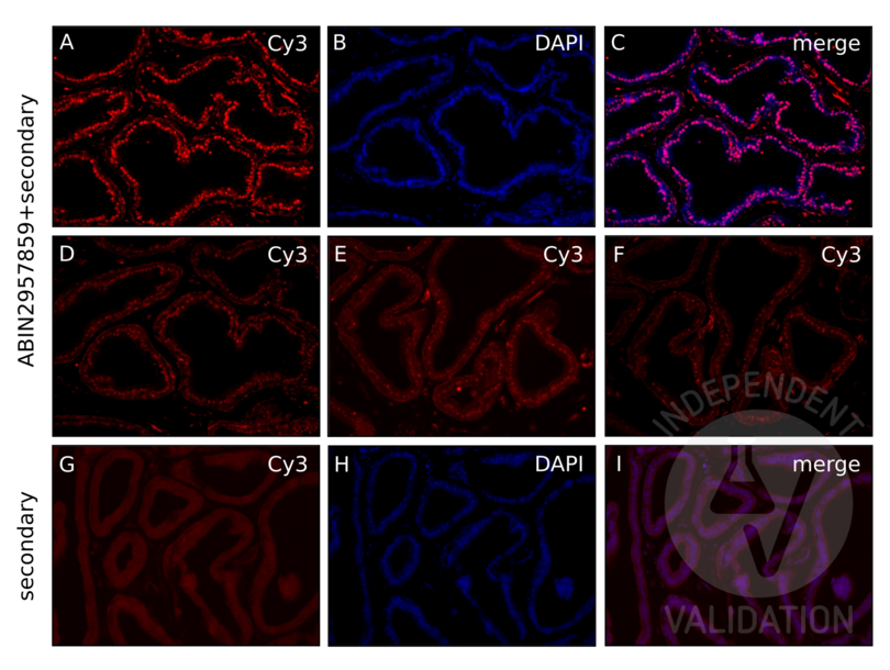
Colocalization of ABIN2957859 staining with the nuclear DAPI stain demonstrates that the AR stain is nuclear. However, there is also some aspecific non-nuclear staining. At 1:200, the staining pattern is similar but weaker. At further dilutions (1:300 and 1:500), the AR staining becomes weaker and the aspecific staining is much stronger. Dilution series was determined by manufacturer's recommendations.
The staining for the AR is clearly nuclear. This is shown by colocalization with the nuclear stain DAPI (B and C). Additionally, the staining pattern seen with the AR antibody is similar to the staining pattern of β-galactosidase seen in the ARlacz transgenic mouse. The ARlacz transgenic mouse expresses β-galactosidase (β-gal) under the control of the AR promoter. Therefore, β-gal is expressed wherever the endogenous AR is expressed.
Exclusion of the primary antibody eliminates nuclear staining, demonstrating that the AR staining is specific. Panels G and H show a prostate that was exposed to secondary antibody, but not primary antibody. The exposure time in Panel G is the same as that for the images shown in the previous figures. The exposure time in Panel H is 5x that shown in the previous figures. Although this isn’t the best negative control to use, we do not have prostate or global AR knockout mice to use for a control.
Validation #101650 (Immunofluorescence)
 Validation Images
![IF staining of prostate from inducible AR-lacZ mouse with ABIN2957859 shows robust staining for the AR at 1:100 dilution (A) and colocalization with nuclear DAPI staining (B, C). Staining becomes weaker and aspecific staining more pronounced at higher dilution factors 1:200, 1:300, and 1:500 (D, E, F). Non-specific signal in the secondary antibody-only control is both extracellular and nuclear. Exposure 5x of that used to image ABIN2957859 was used in G to capture the non-specific signal in the TRITC channel. H shows DAPI imaged at 100ms and I shows that the non-specific signal is both extracellular and nuclear, as shown by the partial colocalization of the non-specific signal with DAPI.]() IF staining of prostate from inducible AR-lacZ mouse with ABIN2957859 shows robust staining for the AR at 1:100 dilution (A) and colocalization with nuclear DAPI staining (B, C). Staining becomes weaker and aspecific staining more pronounced at higher dilution factors 1:200, 1:300, and 1:500 (D, E, F). Non-specific signal in the secondary antibody-only control is both extracellular and nuclear. Exposure 5x of that used to image ABIN2957859 was used in G to capture the non-specific signal in the TRITC channel. H shows DAPI imaged at 100ms and I shows that the non-specific signal is both extracellular and nuclear, as shown by the partial colocalization of the non-specific signal with DAPI.
Protocole
IF staining of prostate from inducible AR-lacZ mouse with ABIN2957859 shows robust staining for the AR at 1:100 dilution (A) and colocalization with nuclear DAPI staining (B, C). Staining becomes weaker and aspecific staining more pronounced at higher dilution factors 1:200, 1:300, and 1:500 (D, E, F). Non-specific signal in the secondary antibody-only control is both extracellular and nuclear. Exposure 5x of that used to image ABIN2957859 was used in G to capture the non-specific signal in the TRITC channel. H shows DAPI imaged at 100ms and I shows that the non-specific signal is both extracellular and nuclear, as shown by the partial colocalization of the non-specific signal with DAPI.
Protocole -
- Format
- Liquid
- Buffer
- Liquid in 0.42 % Potassium phosphate, 0.87 % Sodium chloride, pH 7.3, 30 % glycerol, and 0.01 % sodium azide.
- Agent conservateur
- Sodium azide
- Précaution d'utilisation
- This product contains Sodium azide: a POISONOUS AND HAZARDOUS SUBSTANCE which should be handled by trained staff only.
- Stock
- -20 °C
- Stockage commentaire
- Shipped at 4°C. Upon delivery aliquot and store at -20°C for one year. Avoid freeze/thaw cycles.
- Date de péremption
- 12 months
-
- Antigène
- Androgen Receptor (AR)
- Autre désignation
- Androgen Receptor (AR Produits)
- Synonymes
- anticorps AIS, anticorps DHTR, anticorps HUMARA, anticorps HYSP1, anticorps KD, anticorps NR3C4, anticorps SBMA, anticorps SMAX1, anticorps TFM, anticorps AW320017, anticorps Tfm, anticorps Andr, anticorps AR, anticorps androgen receptor, anticorps AR, anticorps Ar, anticorps ar
- Sujet
- DHTR, NR3C4, Androgen receptor, Dihydrotestosterone receptor, Nuclear receptor subfamily 3 group C member 4
- ID gène
- 367, 11835, 24208
- UniProt
- P10275, P19091, P15207
- Pathways
- Nuclear Receptor Transcription Pathway, Intracellular Steroid Hormone Receptor Signaling Pathway, Steroid Hormone Mediated Signaling Pathway, Regulation of Intracellular Steroid Hormone Receptor Signaling, Nuclear Hormone Receptor Binding, Chromatin Binding
-


