HAVCR1 anticorps (C-Term)
 (1 validation)
-
- Antigène Voir toutes HAVCR1 Anticorps
- HAVCR1 (Hepatitis A Virus Cellular Receptor 1 (HAVCR1))
-
Épitope
- AA 332-348, C-Term
-
Reactivité
- Humain
-
Hôte
- Lapin
-
Clonalité
- Polyclonal
-
Conjugué
- Cet anticorp HAVCR1 est non-conjugé
-
Application
- Western Blotting (WB), Immunocytochemistry (ICC), Immunohistochemistry (Paraffin-embedded Sections) (IHC (p))
- Fonction
- Rabbit IgG polyclonal antibody for Hepatitis A virus cellular receptor 1(HAVCR1) detection. Tested with WB, IHC-P, ICC in Human.
- Séquence
- IKALQNAVEK EVQAEDN
- Réactivité croisée (Details)
- No cross reactivity with other proteins.
- Attributs du produit
-
Rabbit IgG polyclonal antibody for Hepatitis A virus cellular receptor 1(HAVCR1) detection. Tested with WB, IHC-P, ICC in Human.
Gene Name: hepatitis A virus cellular receptor 1
Protein Name: Hepatitis A virus cellular receptor 1(HAVcr-1) - Purification
- Immunogen affinity purified.
- Immunogène
- A synthetic peptide corresponding to a sequence at the C-terminus of human TIM 1(332-348aa IKALQNAVEKEVQAEDN).
- Isotype
- IgG
-
anti-Hepatitis A Virus Cellular Receptor 1 (HAVCR1) (AA 21-240) antibody
HAVCR1 Reactivité: Humain WB, IHC, IP, ICC Hôte: Souris Monoclonal C2 unconjugated
anti-Hepatitis A Virus Cellular Receptor 1 (HAVCR1) (AA 22-235) antibodyHAVCR1 Reactivité: Rat WB, IHC, IP, ICC Hôte: Lapin Polyclonal unconjugated
anti-Hepatitis A Virus Cellular Receptor 1 (HAVCR1) (AA 70-290) antibodyHAVCR1 Reactivité: Humain, Souris WB, ELISA, IHC, ICC, FACS, Neut Hôte: Souris Monoclonal 3D9F5 unconjugated
anti-Hepatitis A Virus Cellular Receptor 1 (HAVCR1) (AA 1-364) antibodyHAVCR1 Reactivité: Humain WB, ELISA, IP Hôte: Souris Monoclonal 2G11 unconjugated
anti-Hepatitis A Virus Cellular Receptor 1 (HAVCR1) (AA 21-240) antibodyHAVCR1 Reactivité: Humain WB, IHC, IP, ICC Hôte: Lapin Polyclonal unconjugated
anti-Hepatitis A Virus Cellular Receptor 1 (HAVCR1) (AA 21-293) antibodyHAVCR1 Reactivité: Humain WB, IHC, IP, ICC Hôte: Lapin Polyclonal unconjugated
anti-Hepatitis A Virus Cellular Receptor 1 (HAVCR1) (AA 22-235) antibodyHAVCR1 Reactivité: Rat WB, IHC, IP, ICC Hôte: Souris Monoclonal C1 unconjugated
anti-Hepatitis A Virus Cellular Receptor 1 (HAVCR1) (AA 21-290) antibodyHAVCR1 Reactivité: Humain WB, ELISA, IHC Hôte: Lapin Polyclonal unconjugated
anti-Hepatitis A Virus Cellular Receptor 1 (HAVCR1) (AA 23-122) antibodyHAVCR1 Reactivité: Humain WB, ELISA Hôte: Souris Polyclonal unconjugated
anti-Hepatitis A Virus Cellular Receptor 1 (HAVCR1) (Center) antibodyHAVCR1 Reactivité: Humain, Souris, Rat WB Hôte: Lapin Polyclonal unconjugated
-
- Indications d'application
-
WB: Concentration: 0.1-0.5 μg/mL, Tested Species: Human
IHC-P: Concentration: 0.5-1 μg/mL, Tested Species: Human, Epitope Retrieval by Heat: Boiling the paraffin sections in 10 mM citrate buffer, pH 6.0, for 20 mins is required for the staining of formalin/paraffin sections.
ICC: Concentration: 0.5-1 μg/mL, Tested Species: Human
Notes: Tested Species: Species with positive results. Predicted Species: Species predicted to be fit for the product based on sequence similarities. Other applications have not been tested. Optimal dilutions should be determined by end users. - Commentaires
-
Antibody can be supported by chemiluminescence kit ABIN921124 in WB, supported by ABIN921231 in IHC(P) and ICC.
- Restrictions
- For Research Use only
-
- by
- Human Protein Atlas
- No.
- #300028
- Date
- 13.12.2016
- Antigène
- HAVCR1
- Numéro du lot
- Application validée
- Immunohistochemistry
- Contrôle positif
- kidney, colon/rectum
- Contrôle négative
- Conclusion
- Passed. ABIN3044226 staining is consistent with RNA-seq data and published literature.
- Anticorps primaire
- ABIN3044226
- Anticorps secondaire
- HRP polymer (ThermoFisher Scientific, TL-125-PH)
- Full Protocol
- Sample preparation:
- Fixation and TMA preparation according to PMID: 22688270.
- Cut 4µm TMA sections using a waterfall microtome (ThermoFisher Scientific, HM355S).
- Dry sections ON at RT and then back them for 12-24h at 50°C.
- Deparaffinization and rehydration in xylene and graded alcohol series:
- xylene twice for 3min.
- 100% ethanol for 1min.
- 95% ethanol for 1min.
- 0.3% H2O2 in 95% ethanol for 5min to block endogenous peroxidase.
- 70% ethanol for 1min.
- 50% ethanol for 1min.
- distilled H2O for 2min.
- Antigen Retrieval by Heat Induced Epitope Retrieval (HIER):
- Boil the sections in Lab Vision Citrate buffer pH6.0 (ThermoFisher Scientific, TA-250-PM1X) for 4min at 125°C in a decloaking chamber (Biocare Medical).
- Allow slides to cool down to 90°C in the decloaking chamber.
- Immunostaining in a Lab Vision Autostainer 480 (ThermorFisher Scientific) at RT with 300µl of reagents for each step:
- Rinse sections in Lab Vision wash buffer with extra tween added to a final concentration of 0.2% (ThermoFisher Scientific, TA-999-TT and TA-125-TW) wash buffer.
- Block sections with Lab Vision Ultra V-Block (ThermoFisher Scientific, TA-125-UB) for 5min.
- Rinse sections 2x in wash buffer.
- Incubate sections with primary rabbit anti-HAVCR1 antibody (antibodies-online, ABIN3044226) diluted 1:2000 for 30min.
- Rinse sections 3x in wash buffer.
- Incubate sections with labeled HRP polymer (ThermoFisher Scientific, TL-125-PH) for 30min.
- Rinse sections 2x in wash buffer.
- Develop in DAB Quanto solution (ThermoFisher Scientific, TL-125-QHDX) for 5min.
- Rinse sections in distilled H2O.
- Washing, counterstaining, and coverslipping in Autostainer XL (Leica Biosystems, ST5010) at RT:
- Counterstaining in Mayer’s hematoxylin plus (HistoLab, 01820) for 7.5min.
- Rinse sections in tap water for 5min.
- Rinse sections in lithium carbonate water diluted 1:5 for 5min.
- Dehydration in graded ethanol series and Neo-Clear (Merck Millipore, 109843) according to the manufacturer recommendations.
- Automated mounting (Leica Biosystems, CV5030) of coverslip with Pertex (Histolab, 00871).
- Microscopy:
- Image acquisition on a Leica Aperio AT2 and Aperio Scanscope AT at 20x magnification.
- Notes
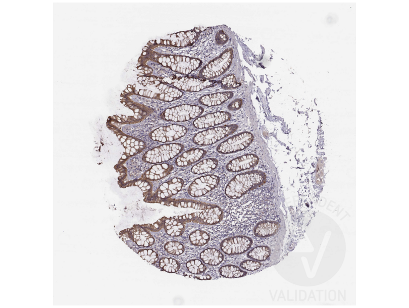
- Based on published literature, HAVCR1 is widely expressed with highest expected levels in kidney and testis. ABIN3044226 stains kidney and colon/rectum where the protein should be enhanced according to RNA-seq data. Staining in kidney is not distinct but the staining is better in colon/rectum.
Validation #300028 (Immunohistochemistry)
 Validation Images
![Staining of human rectum section in a tissue microarray with ABIN3044226. See the protocol for detailed information.]() Staining of human rectum section in a tissue microarray with ABIN3044226. See the protocol for detailed information.
Protocole
Staining of human rectum section in a tissue microarray with ABIN3044226. See the protocol for detailed information.
Protocole -
- Format
- Lyophilized
- Reconstitution
- Add 0.2 mL of distilled water will yield a concentration of 500 μg/mL.
- Concentration
- 500 μg/mL
- Buffer
- Each vial contains 5 mg BSA, 0.9 mg NaCl, 0.2 mg Na2HPO4, 0.05 mg Thimerosal, 0.05 mg Sodium azide.
- Agent conservateur
- Thimerosal (Merthiolate), Sodium azide
- Précaution d'utilisation
- This product contains Sodium azide and Thimerosal (Merthiolate): POISONOUS AND HAZARDOUS SUBSTANCES which should be handled by trained staff only.
- Conseil sur la manipulation
- Avoid repeated freezing and thawing.
- Stock
- 4 °C/-20 °C
- Stockage commentaire
-
At -20°C for one year. After reconstitution, at 4°C for one month.
It can also be aliquotted and stored frozen at -20 °C for a longer time. Avoid repeated freezing and thawing. - Date de péremption
- 12 months
-
- Antigène
- HAVCR1 (Hepatitis A Virus Cellular Receptor 1 (HAVCR1))
- Autre désignation
- HAVCR1 (HAVCR1 Produits)
- Synonymes
- anticorps HAVCR, anticorps HAVCR-1, anticorps KIM-1, anticorps KIM1, anticorps TIM, anticorps TIM-1, anticorps TIM1, anticorps TIMD-1, anticorps TIMD1, anticorps Kim1, anticorps HAVCR1, anticorps LOC100226241, anticorps AI503787, anticorps Tim1, anticorps Timd1, anticorps hepatitis A virus cellular receptor 1, anticorps hepatitis A virus cellular receptor 1 homolog, anticorps HAVCR1, anticorps Havcr1, anticorps LOC100226241
- Classe de substances
- Virus
- Sujet
-
KIM1(KIDNEY INJURY MOLECULE 1), also known as HAVCR1, HAVCR or TIM1, is a protein that in humans is encoded by the KIM1 gene. The KIM1 gene is mapped to 5q33.3. Biochemical, mutational, and cell adhesion analyses confirm that Tim1 is capable of homophilic Tim-Tim interactions. The features identified in murine KIM1 is conserved in human KIM1. The KIM1 protein is indeed a receptor for the virus through the infection of canine osteogenic sarcoma cells expressing HAVCR1 with HAV. Using a monoclonal antibody to mouse Tim1, Tim1 is expressed after activation of naive T cells and on T cells differentiated in Th2-polarizing conditions. Ectopic expression of KIM1 during mouse T-cell differentiation leads to production of the Th2-type cytokine Il4, but not the Th1-type cytokine Ifng. KIM1-expressing epithelial cells internalized apoptotic bodies, and Kim1 is directly responsible for phagocytosis in cultured primary rat tubule epithelial cells and in porcine and canine epithelial cell lines.
Synonyms: HAVCR 1 antibody|HAVcr-1 antibody|HAVCR1 antibody|Hepatitis A virus cellular receptor 1 antibody|Kidney injury molecule 1 antibody|KIM 1 antibody|KIM-1 antibody|T cell immunoglobin domain and mucin domain protein 1 antibody|T-cell immunoglobulin and mucin domain-containing protein 1 antibody|T-cell membrane protein 1 antibody|TIM antibody|TIM-1 antibody|TIM1 antibody|TIMD 1 antibody|TIMD-1 antibody|TIMD1 antibody|TIMD1_HUMAN antibody - UniProt
- Q96D42
-


