COL3 anticorps
 (5 références)
 (1 validation)
-
- Antigène Voir toutes COL3 Anticorps
- COL3 (Collagen, Type III (COL3))
-
Reactivité
- Humain, Boeuf (Vache), Mammifères
-
Hôte
- Lapin
-
Clonalité
- Polyclonal
-
Conjugué
- Cet anticorp COL3 est non-conjugé
-
Application
- Western Blotting (WB), ELISA, Immunohistochemistry (IHC), Immunoprecipitation (IP)
- Réactivité croisée (Details)
- Typically negligible cross-reactivity against other types of collagens was detected by ELISA against purified standards. Some class-specific anti-collagens may be specific for three-dimensional epitopes which may result in diminished reactivity with denatured collagen or formalin-fixed, paraffin embedded tissues. This antibody reacts with most mammalian Type III collagens and has negligible cross-reactivity with Type I, II, IV, V or VI collagens. Non-specific cross-reaction of anti-collagen antibodies with other human serum proteins or non-collagen extracellular matrix proteins is negligible.
- Purification
- Collagen III Antibody has been prepared by immunoaffinity chromatography using immobilized antigens followed by extensive cross-adsorption against other collagens, human serum proteins and non-collagen extracellular matrix proteins to remove any unwanted specificities.
- Immunogène
-
Immunogen: Collagen Type III from human and bovine placenta
Immunogen Type: Native Protein
- Isotype
- IgG
-
anti-Collagen, Type III (COL3) (AA 1301-1400) antibody
COL3 Reactivité: Humain, Rat, Souris, Lapin, Chien WB, ELISA, IHC (fro), IF (cc), IF (p), IHC (p) Hôte: Lapin Polyclonal unconjugated
anti-Collagen, Type III (COL3) (AA 801-900) antibodyCOL3 Reactivité: Humain, Rat, Souris WB, ELISA, IHC (fro), IF (cc), IF (p), IHC (p) Hôte: Lapin Polyclonal unconjugated
anti-Collagen, Type III (COL3) antibodyCOL3 Reactivité: Humain WB, IF, IP Hôte: Lapin Monoclonal unconjugated
anti-Collagen, Type III (COL3) antibodyCOL3 Reactivité: Humain ELISA Hôte: Chèvre Polyclonal unconjugated
anti-Collagen, Type III (COL3) antibody (HRP)COL3 Reactivité: Humain, Boeuf (Vache), Mammifères WB, ELISA, IHC, IP Hôte: Lapin Polyclonal HRP
anti-Collagen, Type III (COL3) antibody (Biotin)COL3 Reactivité: Humain ELISA Hôte: Chèvre Polyclonal Biotin
anti-Collagen, Type III (COL3) antibodyCOL3 Reactivité: Humain, Rat, Souris WB Hôte: Souris Monoclonal Q76 unconjugated
anti-Collagen, Type III (COL3) antibody (Biotin)COL3 Reactivité: Humain, Boeuf (Vache), Mammifères WB, ELISA, IHC, IP Hôte: Lapin Polyclonal Biotin
anti-Collagen, Type III (COL3) antibody (FITC)COL3 Reactivité: Humain, Boeuf (Vache), Mammifères WB, ELISA, IHC, IP Hôte: Lapin Polyclonal FITC
anti-Collagen, Type III (COL3) antibody (Magnetic Particles)COL3 Reactivité: Humain, Rat, Souris IP Hôte: Souris Monoclonal Col-29 Magnetic Particles
-
- Indications d'application
-
Immunohistochemistry Dilution: 1:50 - 1:200
Application Note: Anti-Collagen antibodies have been used for indirect trapping ELISA for quantitation of antigen in serum using a standard curve, immunoprecipitation, native (non-denaturing, non-dissociating) PAGE, immunohistochemistry, and western blotting for highly sensitive qualitative analysis.
Western Blot Dilution: 1:1,000 - 1:10,000
Immunoprecipitation Dilution: 1:100
ELISA Dilution: 1:5,000 - 1:50,000
- Restrictions
- For Research Use only
-
- by
- MS Validated Antibodies
- No.
- #300050
- Date
- 22.08.2023
- Antigène
- COL3
- Numéro du lot
- 47783
- Application validée
- Immunohistochemistry
- Contrôle positif
Human TMA
Mouse monoclonal anti-COL3 antibody clone FH-7A
rabbit monoclonal anit-COL3 antibody HL1906
- Contrôle négative
- Conclusion
Passed. The anti-COL3 antibody ABIN5596830 stains collagen III in formalin-fixed tissues consistently with expected staining pattern.
- Anticorps primaire
- ABIN5596830
- Anticorps secondaire
- EnVision Polymer-HRP mouse/rabbit Kit, Dako REAL, K5007
- Full Protocol
- Slide preparation
- Mount 2,5 µm FFPE tissue sections on superfrost slides.
- Deparaffinize tissue sections 3x 5 min in xylene.
- Rehydrate tissue sections in a descending ethanol series for 1 min each 100%, 96%, and 80% ethanol.
- Rinse tissue sections for 5 min in TBST buffer (DAKO, K8000).
- Epitope retrieval
- Autoclave tissue sections for 5 min at 121 °C in 1x Tris-EDTA-citrate buffer pH7.8 (20x Tris-EDTA-citrate buffer stock solution: 5 g Trizma base (Sigma-Aldrich, T1503), 10 g EDTA (Merck, 1.08418), 6.4g tri-sodium citrate (Sigma-Aldrich, C0909), adjust to pH 7.8 using HCL 1 M, ad 1 L with dH2O).
- Rinse tissue sections for 5 min in TBST buffer.
- Peroxidase blocking
- Incubate tissue sections for 10 min in Peroxidase-Blocking Solution (Dako REAL, S2023).
- Rinse tissue sections 2x for 5 min in TBST buffer.
- Antibody incubation
- o Dilute primary rabbit anti-collagen III antibody (antibodies-online, ABIN5596830, lot 47783) diluted 1:112.5, 1:225, or 1:450 in antibody diluent (Dako REAL, S2022).
- Cover tissue section with 100-200 µl diluted antibody.
- Incubate tissue sections for 1 h at 37 °C in a moist chamber.
- Rinse tissue sections for 5 min in TBST buffer.
- Apply EnVision Polymer-HRP mouse/rabbit Kit (Dako REAL, K5007) according to manufacturer’s recommendation.
- Rinse tissue sections 2x for 5 min in TBST buffer.
- Staining
- Cover slides for 10 min with DAB-Chromogen (EnVision Polymer-HRP mouse/rabbit Kit, Dako REAL, K5007).
- Wash slides thoroughly with dH2O.
- Counterstain for 15 sec with Hematoxylin (Mayers Hematoxylin: 200ml ddH2O, 0,2g Hematoxylin (Serva, 24420.02), 10 g aluminium potassium sulfate dodecahydrate (Merck, 1.01047), 0,04 g sodium iodate (Merck, 1.06525), 10 g chloral hydrate (Sigma-Aldrich, 15307)).
- Develop for 15 sec in H2O.
- Dehydrate tissue sections in an ascending ethanol series for 1 min each 80%, 96%, 100% ethanol.
- Wash tissue sectiona 3x 5 min in xylene.
- Apply mounting medium and coverslips.
- Image acquisition
- o Acquire images using a Galileo TMAtic (ISENET).
- Notes
For antibody comparison an antibody test TMA was used that contained 80 normal tissues from 21 different organs and 95 neoplastic tissues from 18 different tumor types.
The anti-COL3 antibody ABIN5596830 tends to cause a rather ubiquitous cytoplasmic staining of all kinds of cells including epithelial cells. This cytoplasmic staining is reduced to a tolerable level at dilutions of 1:225 and less. At such dilutions, ABIN5596830 stains fibrillar structures – also detected by the two reference antibodies - at weak to moderate intensity. Based on the identical staining pattern obtained by three different collagen III antibodies, these fibrillar staining’s are likely due to a binding of the antibodies to collagen III. Collagen III staining is typically located adjacent to epithelial structures, in smooth muscles or around vessels of all sizes, and in the stroma of tumors.
ABIN5596830 cross-reacts with some inflammatory cells in lymph nodes.
Validation #300050 (Immunohistochemistry)
 Validation Images
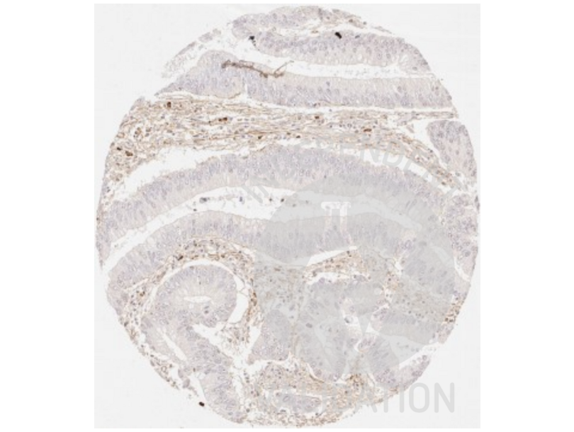
![IHC staining of the stroma in colorectal cancer with anti-collagen III antibody ABIN5596830 diluted 1:225 shows fibrillar collagen III staining.]() IHC staining of the stroma in colorectal cancer with anti-collagen III antibody ABIN5596830 diluted 1:225 shows fibrillar collagen III staining.
IHC staining of the stroma in colorectal cancer with anti-collagen III antibody ABIN5596830 diluted 1:225 shows fibrillar collagen III staining.
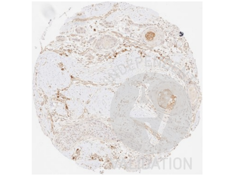
![IHC staining of squamous cell carcinoma of the oral cavity with anti-collagen III antibody ABIN5596830 diluted 1:225 shows a distinct fibrillar collagen III staining of the stroma.]() IHC staining of squamous cell carcinoma of the oral cavity with anti-collagen III antibody ABIN5596830 diluted 1:225 shows a distinct fibrillar collagen III staining of the stroma.
IHC staining of squamous cell carcinoma of the oral cavity with anti-collagen III antibody ABIN5596830 diluted 1:225 shows a distinct fibrillar collagen III staining of the stroma.
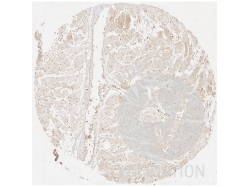
![IHC staining of heart tissue with anti-collagen III antibody ABIN5596830 diluted 1:225 shows distinct fibrillar collagen III staining surrounding each hear muscle cell.]() IHC staining of heart tissue with anti-collagen III antibody ABIN5596830 diluted 1:225 shows distinct fibrillar collagen III staining surrounding each hear muscle cell.
IHC staining of heart tissue with anti-collagen III antibody ABIN5596830 diluted 1:225 shows distinct fibrillar collagen III staining surrounding each hear muscle cell.
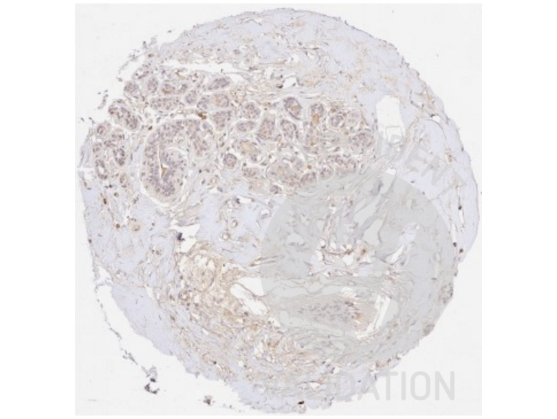
![IHC staining of non-cancerous breast tissue with anti-collagen III antibody ABIN5596830 diluted 1:225 shows considerable sclerosis.]() IHC staining of non-cancerous breast tissue with anti-collagen III antibody ABIN5596830 diluted 1:225 shows considerable sclerosis.
Protocole
IHC staining of non-cancerous breast tissue with anti-collagen III antibody ABIN5596830 diluted 1:225 shows considerable sclerosis.
Protocole -
- Format
- Liquid
- Concentration
- 1.0 mg/mL
- Buffer
-
Buffer: 0.125 M Sodium Borate, 0.075 M Sodium Chloride, 0.005 M EDTA, pH 8.0
Stabilizer: None
- Agent conservateur
- Sodium azide
- Précaution d'utilisation
- This product contains Sodium azide: a POISONOUS AND HAZARDOUS SUBSTANCE which should be handled by trained staff only.
- Stock
- 4 °C,-20 °C
- Stockage commentaire
- Store vial at 4° C prior to opening. This product is stable at 4° C as an undiluted liquid. Dilute only prior to immediate use. For extended storage, mix with an equal volume of glycerol, aliquot contents and freeze at -20° C or below. Avoid cycles of freezing and thawing.
- Date de péremption
- 12 months
-
-
: "Keloid Pathogenesis: Potential Role of Cellular Fibronectin with the EDA Domain." dans: The Journal of investigative dermatology, Vol. 135, Issue 7, pp. 1921-1924, (2015) (PubMed).
: "Overexpression of Twinkle-helicase protects cardiomyocytes from genotoxic stress caused by reactive oxygen species." dans: Proceedings of the National Academy of Sciences of the United States of America, Vol. 110, Issue 48, pp. 19408-13, (2014) (PubMed).
: "Propolis Modifies Collagen Types I and III Accumulation in the Matrix of Burnt Tissue." dans: Evidence-based complementary and alternative medicine : eCAM, Vol. 2013, pp. 423809, (2013) (PubMed).
: "The role of decorin and biglycan dermatan sulfate chain(s) in fibrosis-affected fascia." dans: Glycobiology, Vol. 21, Issue 10, pp. 1301-16, (2012) (PubMed).
: "Genetic deletion of the angiotensin-(1-7) receptor Mas leads to glomerular hyperfiltration and microalbuminuria." dans: Kidney international, Vol. 75, Issue 11, pp. 1184-1193, (2009) (PubMed).
-
: "Keloid Pathogenesis: Potential Role of Cellular Fibronectin with the EDA Domain." dans: The Journal of investigative dermatology, Vol. 135, Issue 7, pp. 1921-1924, (2015) (PubMed).
-
- Antigène
- COL3 (Collagen, Type III (COL3))
- Autre désignation
- Collagen Type III (COL3 Produits)
- Synonymes
- anticorps PT, anticorps RPRGL2, anticorps THPH1, anticorps Cf-2, anticorps Cf2, anticorps FII, anticorps thrombin, anticorps prothrombin, anticorps coagulation factor II, thrombin, anticorps coagulation factor II, anticorps F2
- Sujet
-
Synonyms: Collagen type III alpha 1 antibody, Collagen type III alpha antibody, EDS4A antibody, Ehlers Danlos syndrome type IV, autosomal dominant antibody, Fetal collagen antibody, COL3A1
Background: Rockland produces highly active antibodies and conjugates to collagens. Collagens are highly conserved throughout evolution and are characterized by an uninterrupted ''Glycine-X-Y'' triplet repeat that is a necessary part of the triple helical structure. For these reasons, it is often extremely difficult to generate antibodies with specificities to collagens. The development of 'type' specific antibodies is dependent on NON-DENATURED three-dimensional epitopes. Rockland extensively purifies collagens for immunization from human and bovine placenta and cartilage by limited pepsin digestion and selective salt precipitation. This preparation results in a native conformation of the protein. Antibodies are isolated from rabbit antiserum and are extensively cross-adsorbed by immunoaffinity purification to produce 'type' specific antibodies. Greatly diminished reactivity and selectivity of these antibodies will result if denaturing and reducing conditions are used for SDS-PAGE and immunoblotting. Ideal for investigators involved in Cell Biology, Signal Transduction and Stem Cell research.
Gene Name: COL3A1
- ID gène
- 1281
- UniProt
- P02461
-


