BIN1 Protein (Transcript Variant 8) (Myc-DYKDDDDK Tag)
 (1 validation)
-
- Antigène Voir toutes BIN1 Protéines
- BIN1 (Bridging Integrator 1 (BIN1))
- Type de proteíne
- Recombinant
- Attributs du protein
- Transcript Variant 8
-
Origine
- Humain
-
Source
- HEK-293 Cells
- Purification/Conjugué
- Cette BIN1 protéine est marqué à la Myc-DYKDDDDK Tag.
- Application
- Antibody Production (AbP), Standard (STD)
- Attributs du produit
-
- Recombinant human BIN1 / AMPHL (transcript variant 8) protein expressed in HEK293 cells.
- Produced with end-sequenced ORF clone
- Pureté
- > 80 % as determined by SDS-PAGE and Coomassie blue staining
-
Bridging Integrator 1 (BIN1) (AA 355-454) protein (GST tag)
BIN1 Origine: Humain Hôte: Wheat germ Recombinant WB, ELISA, AA, AP
Bridging Integrator 1 (BIN1) (AA 1-439) protein (GST tag)BIN1 Origine: Humain Hôte: Wheat germ Recombinant WB, ELISA, AA, AP
Bridging Integrator 1 (BIN1) (AA 2-593) protein (His tag)Crystallography grade BIN1 Origine: Humain Hôte: Cellules d'insectes Recombinant >95 % as determined by SDS PAGE, Size Exclusion Chromatography and Western Blot. SDS, WB, ELISA, Crys
Bridging Integrator 1 (BIN1) (AA 2-588) protein (His tag)Crystallography grade BIN1 Origine: Souris Hôte: Cellules d'insectes Recombinant >95 % as determined by SDS PAGE, Size Exclusion Chromatography and Western Blot. SDS, WB, ELISA, Crys
Bridging Integrator 1 (BIN1) (AA 1-439) protein (His tag)BIN1 Origine: Humain Hôte: Escherichia coli (E. coli) Recombinant > 90 % by SDS – PAGE SDS
Bridging Integrator 1 (BIN1) (Transcript Variant 9) protein (Myc-DYKDDDDK Tag)BIN1 Origine: Humain Hôte: HEK-293 Cells Recombinant > 80 % as determined by SDS-PAGE and Coomassie blue staining AbP, STD
Bridging Integrator 1 (BIN1) (Transcript Variant 2) protein (Myc-DYKDDDDK Tag)BIN1 Origine: Humain Hôte: HEK-293 Cells Recombinant > 80 % as determined by SDS-PAGE and Coomassie blue staining AbP, STD
-
- Indications d'application
-
Recombinant human proteins can be used for:
Native antigens for optimized antibody production
Positive controls in ELISA and other antibody assays - Commentaires
-
The tag is located at the C-terminal.
- Restrictions
- For Research Use only
-
- by
- Department of Medical Physiology, UMC Utrecht
- No.
- #102757
- Date
- 03.05.2018
- Antigène
- BIN1
- Numéro du lot
- 10C425
- Application validée
- Western Blotting
- Contrôle positif
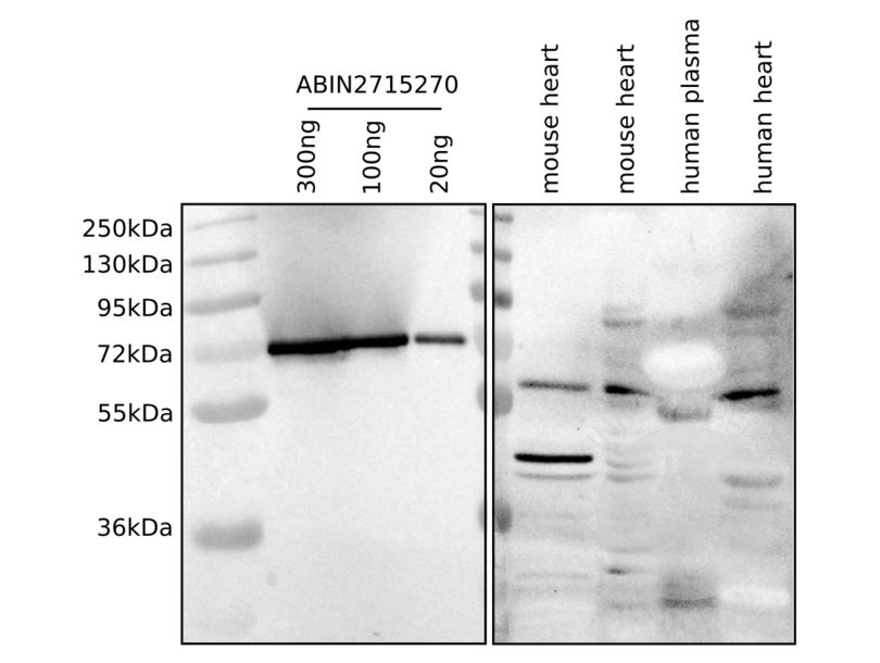
- BIN1 antibody, endogenous BIN1 in mouse heart lysates and human plasma and heart lysate
- Contrôle négative
- none
- Conclusion
Passed. The recombinant BIN1 protein ABIN2715270 works excellently in western blot and appears at the expected MW.
- Anticorps primaire
- rabbit anti-BIN1 antibody (Sigma-Aldrich, HPA003894, lot R04603)
- Anticorps secondaire
- goat anti-rabbit HRP-conjugated antibody (Bio-Rad Cat, 170-6515)
- Full Protocol
- Lyse half a murine heart or a corresponding piece (in size) of human heart tissue in 250µl of lysis buffer (20mM Tris-HCl pH7.4, 150mM NaCl, 10mM Na2HPO4, 1% Triton X-100, 1% Na-deoxycholate, 0.1% SDS, 1mM EDTA, 50mM NaF).
- Determine total protein content of the lysates and human plasma samples using Pierce BCA Protein Assay Kit (ThermoFisher Scientific, 23227, lot SC246925).
- Sample preparation:
- Dilute appropriate amount of lysate in lysis buffer + 5x Laemmli SDS sample buffer (lysis buffer:sample buffer 4:1) to achieve 25µg of protein per 20µl of sample, boil the samples for 5min at 96°C.
- Dilute appropriate amount of human plasma in lysis buffer + 5x Laemmli SDS sample buffer (lysis buffer:sample buffer 4:1) to achieve 25µg of protein per 20µl of sample, boil the samples for 5min at 96°C.
- 20ng, 100ng, and 300ng recombinant BIN1 (antibodies-online, ABIN2715270, lot 10C425) were diluted in 20µl ddH2O + 5x Laemmli SDS sample buffer (ddH2O:sample buffer 4:1), samples were boiled for5min at 96°C.
- Separate the samples on a freshly cast discontinuous denaturing SDS-PAGE 10% running gel in an electrophoresis chamber (Bio-Rad) for approximately 20min at 75-100V for the stacking and 1h at 100-200V for the running gel.
- Transfer proteins onto a nitrocellulose membrane 0.45µm (Bio-Rad, 1620115, lot A10206645) with a Hoefer TE 77 semi-dry transfer unit (Amersham Biosciences) for 70min at 43mA/gel.
- Verify protein transfer through Ponceau Red staining.
- Block the membrane with blocking buffer (TBST containing 5% BSA) for 1h at RT.
- Incubation with primary rabbit anti-BIN1 antibody (Sigma-Aldrich, HPA003894, lot R04603) diluted 1:500 in blocking buffer for ON at 4°C.
- Wash membrane 3x for 10min with TBST.
- Incubation with secondary goat anti-rabbit HRP-conjugated antibody (Bio-Rad Cat, 170-6515) diluted 1:7000 in blocking buffer for 1-2h at RT.
- Wash membrane 3x for 10min with TBST.
- Reveal protein bands using ECL substrate (GE Healthcare, RPN2232, lot 13601176) on an ChemiDoc XRS + (BioRad) and ImageLab (version 6.0.0 build 25, standard edition. 2017, BioRad laboratories, Inc) software was used to determine best exposure time for optimal visualization of bands.
- Notes
The BIN1 antibody reveals a protein of the expected MW. ABIN2715270 migrates higher than the endogenous murine and human proteins due to the C-terminal myc- and DYKDDDDK-tags.
Validation #102757 (Western Blotting)
 Validation Images
![Western blot analysis of human recombinant BIN1 ABIN2715270 (left panel) in comparison to the endogenous murine and human proteins (right panel).]() Western blot analysis of human recombinant BIN1 ABIN2715270 (left panel) in comparison to the endogenous murine and human proteins (right panel).
Protocole
Western blot analysis of human recombinant BIN1 ABIN2715270 (left panel) in comparison to the endogenous murine and human proteins (right panel).
Protocole -
- Concentration
- 50 μg/mL
- Buffer
- 25 mM Tris.HCl, pH 7.3, 100 mM glycine, 10 % glycerol.
- Stock
- -80 °C
- Stockage commentaire
- Store at -80°C. Thaw on ice, aliquot to individual single-use tubes, and then re-freeze immediately. Only 2-3 freeze thaw cycles are recommended.
-
- Antigène
- BIN1 (Bridging Integrator 1 (BIN1))
- Autre désignation
- Bin1,amphl (BIN1 Produits)
- Synonymes
- bin1 Protein, MGC53185 Protein, amph2 Protein, amphl Protein, sh3p9 Protein, MGC76187 Protein, cb57 Protein, zgc:86701 Protein, BIN1 Protein, AMPH2 Protein, AMPHL Protein, SH3P9 Protein, ALP-1 Protein, Amphl Protein, BRAMP-2 Protein, bridging integrator 1 Protein, bridging integrator 1 S homeolog Protein, bridging integrator 1b Protein, BIN1 Protein, bin1.S Protein, bin1 Protein, bin1b Protein, Bin1 Protein
- Sujet
- This gene encodes several isoforms of a nucleocytoplasmic adaptor protein, one of which was initially identified as a MYC-interacting protein with features of a tumor suppressor. Isoforms that are expressed in the central nervous system may be involved in synaptic vesicle endocytosis and may interact with dynamin, synaptojanin, endophilin, and clathrin. Isoforms that are expressed in muscle and ubiquitously expressed isoforms localize to the cytoplasm and nucleus and activate a caspase-independent apoptotic process. Studies in mouse suggest that this gene plays an important role in cardiac muscle development. Alternate splicing of the gene results in several transcript variants encoding different isoforms. Aberrant splice variants expressed in tumor cell lines have also been described.
- Poids moléculaire
- 50 kDa
- NCBI Accession
- NP_004296
-


