ODF2 anticorps
 (2 références)
 (2 validations)
Aperçu rapide pour ODF2 anticorps (ABIN2430583)
Antigène
Voir toutes ODF2 AnticorpsReactivité
Hôte
Clonalité
Conjugué
Application
-
-
Purification
- Affinity purification
-
Immunogène
- Recombinant protein of human ODF2
-
Isotype
- IgG
-
-
anti-Outer Dense Fiber of Sperm Tails 2 (ODF2) (AA 706-804) antibody
ODF2 Reactivité: Humain IF Hôte: Souris Monoclonal 1A1 unconjugated
anti-Outer Dense Fiber of Sperm Tails 2 (ODF2) (AA 630-829) antibodyODF2 Reactivité: Humain WB, IF Hôte: Lapin Polyclonal unconjugated
anti-Outer Dense Fiber of Sperm Tails 2 (ODF2) (Internal Region) antibodyVerified ODF2 Reactivité: Souris WB, ELISA Hôte: Chèvre Polyclonal unconjugated
anti-Outer Dense Fiber of Sperm Tails 2 (ODF2) (AA 218-267) antibodyODF2 Reactivité: Humain, Souris, Rat, Chien, Cobaye, Cheval, Boeuf (Vache), Lapin, Porc, Singe, Roussette (Chauve-souris), Poulet WB Hôte: Lapin Polyclonal unconjugated
anti-Outer Dense Fiber of Sperm Tails 2 (ODF2) (N-Term) antibodyODF2 Reactivité: Humain, Souris, Rat, Chien, Cobaye, Cheval, Boeuf (Vache), Lapin, Porc, Singe, Roussette (Chauve-souris) WB Hôte: Lapin Polyclonal unconjugated
anti-Outer Dense Fiber of Sperm Tails 2 (ODF2) (N-Term) antibodyODF2 Reactivité: Humain, Souris, Rat WB Hôte: Lapin Polyclonal unconjugated
anti-Outer Dense Fiber of Sperm Tails 2 (ODF2) antibodyODF2 Reactivité: Humain WB Hôte: Lapin Monoclonal unconjugated
-
-
Indications d'application
- WB 1:500-1:2000, IHC 1:25-1:100
-
Restrictions
- For Research Use only
-
-
- by
- Georg-August-University of Göttingen, Johann-Friedrich-Blumenbach-Institute for Zoology and Anthropology, Developmental Biology
- No.
- #100023
- Date
- 03.05.2016
- Antigène
- Outer Dense Fiber of Sperm Tails 2 (ODF2)
- Numéro du lot
- Application validée
- Western Blotting
- Contrôle positif
- recombinant ODF2-His, bacterially expressed; NIH3T3 total cell lysate
- Contrôle négative
- Conclusion
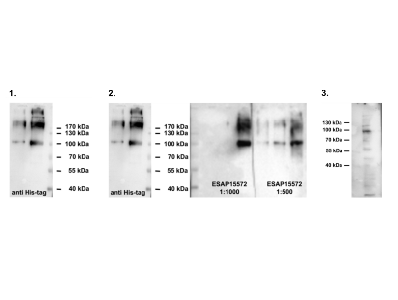
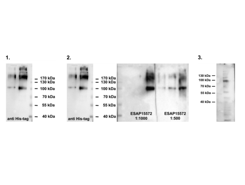
- Passed, regarding sensitivity and specificity. The antibody detects bacterially expressed ODF2 (see 2)) and the endogenous protein (see 3)) with only moderate background staining in WB.
- Anticorps primaire
- ABIN2430582
- Anticorps secondaire
- anti rabbit IgG (H+L), HRP-linked (Jackson Immuno Research, Code number: 111-035-003, Lot number: 123450)
- Full Protocol
- Proteins separated on 10% SDS-PAGE (Laemmli 1970), blotted to Protran (GE Healthcare No. 10600004, 0.2 µm) (Towbin et al., 1979). Blocking of membrane in 5% skim milk in TBST (50 mM Tris-HCl, pH 7.4, 150 mM NaCl, 0.1% Tween 20) for 30 min at room temperature.
- Incubation with first antibody in 5% skim milk in TBST at 4°C overnight.
- Washing in TBST for 30 minutes at room temperature.
- Incubation with secondary antibody in 5% skim milk in TBST for 45 minutes at room temperature. Washing in TBST for 30-45 minutes at room temperature.
- Secondary antibody: anti rabbit IgG (H+L), HRP-linked (Jackson Immuno Research, Code number: 111-035-003, Lot number: 123450). Dilution: 1:10,000.
- Chemiluminescence detection (Western Lightning Ultra, Lot 215-15091, Perkin Elmer) and image capture via BioRad Chemdoc or X-ray films.
- Notes
Validation #100023 (Western Blotting)
 Validation Images
![1. Positive control: protein ODF2-His, bacterially expressed, different amounts per lane, detection with anti His-Tag antibody (Novagen, 70796-3, Lot number N37164) (1:1,000). Secondary antibody anti mouse-HRP (Sigma-Aldrich A9044, Batch Number 034M4761) (1:10,000). 2. Test: protein ODF2-His, bacterially expressed, different amounts per lane, detection with anti ODF2 (ABIN2430582). 3.NIH3T3 total cell lysate (125µg/per lane), detection with anti ODF2 (ABIN2430582). Expected molecular weight of endogenous ODF2]() 1. Positive control: protein ODF2-His, bacterially expressed, different amounts per lane, detection with anti His-Tag antibody (Novagen, 70796-3, Lot number N37164) (1:1,000). Secondary antibody anti mouse-HRP (Sigma-Aldrich A9044, Batch Number 034M4761) (1:10,000). 2. Test: protein ODF2-His, bacterially expressed, different amounts per lane, detection with anti ODF2 (ABIN2430582). 3.NIH3T3 total cell lysate (125µg/per lane), detection with anti ODF2 (ABIN2430582). Expected molecular weight of endogenous ODF2
Protocole
1. Positive control: protein ODF2-His, bacterially expressed, different amounts per lane, detection with anti His-Tag antibody (Novagen, 70796-3, Lot number N37164) (1:1,000). Secondary antibody anti mouse-HRP (Sigma-Aldrich A9044, Batch Number 034M4761) (1:10,000). 2. Test: protein ODF2-His, bacterially expressed, different amounts per lane, detection with anti ODF2 (ABIN2430582). 3.NIH3T3 total cell lysate (125µg/per lane), detection with anti ODF2 (ABIN2430582). Expected molecular weight of endogenous ODF2
Protocole -
- by
- Georg-August-University of Göttingen, Johann-Friedrich-Blumenbach-Institute for Zoology and Anthropology, Developmental Biology
- No.
- #100024
- Date
- 03.05.2016
- Antigène
- Outer Dense Fiber of Sperm Tails 2 (ODF2)
- Numéro du lot
- Application validée
- Immunocytochemistry
- Contrôle positif
- mouse anti-γ-tubulin (Sigma-Aldrich, clone GTU-88, T6557, lot number 072M4808)
- Contrôle négative
- Conclusion
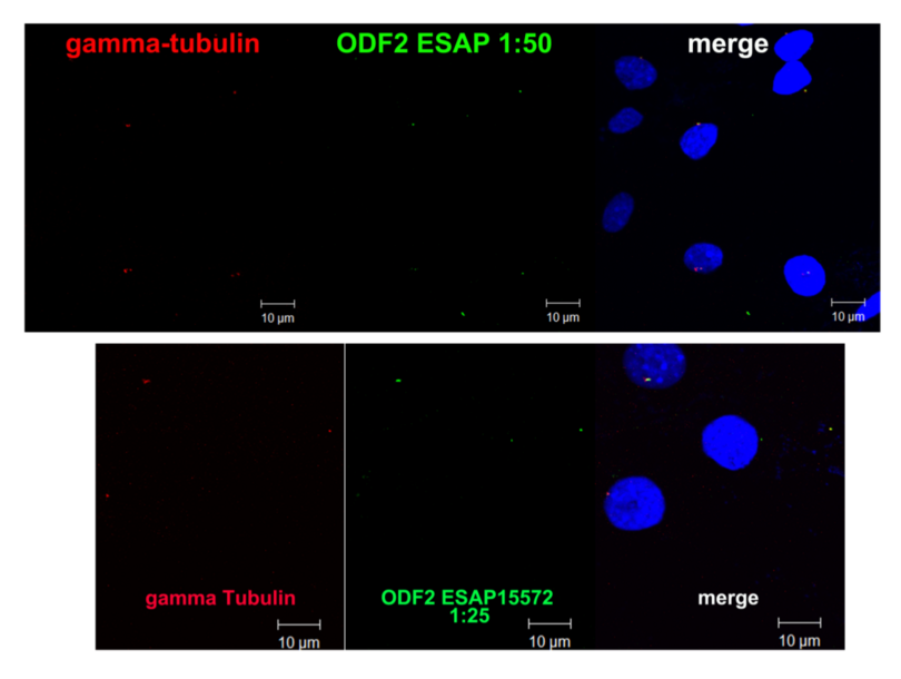
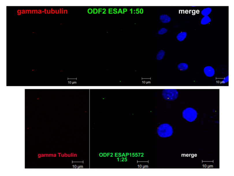
- Passed, the antibody detects centrosomal ODF2 in immunocytological preparations with only very low background staining (the green dots are mostly the correct location of ODF2 as they overlap with γ-tubulin spots (in red)).
- Anticorps primaire
- ABIN2430582
- Anticorps secondaire
- MFP488 goat anti-rabbit IgG (H+L) (MoBiTec, #MFP-A1008)
- Full Protocol
- NIH3T3 cells (ATCC) were grown on cover slips in DMEM, 10% fetal bovine serum, 5% penicillin/streptomycin (all Gibco) at 37°C in 5% CO2.
- Cells were fixed in ice-cold methanol for 20 min at -20°C.
- Unspecific binding sites were blocked in PBST (phosphate buffered saline (PBS) containing 0.15% bovine serum albumin, 0.1% Tween-20) for 1 hr.
- Cells were incubated with the primary antibodies at 4°C overnight.
- Cells were washed in TBST (50 mM Tris-HCl, pH 7.4, 150 mM NaCl, 0.1% Tween 20) for 15 min.
- Cells were incubated with secondary antibodies and DAPI for 1h at 37°C.
- Cells were washed in TBST for 15 min.
- Cells were embedded in Dako Fluorescent Mounting Medium (Dako).
- As positive control for centrosomal decoration anti ?-tubulin (Sigma-Aldrich, clone GTU-88, T6557, lot number 072M4808; 1:100) was used. anti ?-tubulin was detected with MFP590 goat anti-mouse IgG (H+L) (MoBiTec, #MFP-A1032; 1:300).
- Anti ODF2-antibody was detected with MFP488 goat anti-rabbit IgG (H+L) (MoBiTec, #MFP-A1008; 1:300).
- DNA was counterstained with DAPI.
- Images were taken by confocal microscopy (LSM 510, Zeiss), and processed using Adobe Photoshop 5.0.
- Notes
Validation #100024 (Immunocytochemistry)
 Validation Images
![Anti-ODF2 antibody ABIN2430582 (ESAP15572) is detected with a MFP488 goat anti-rabbit IgG (H+L) secondary antibody (green). The control mouse anti-γ-tubulin antibody is detected with a MFP590 goat anti-mouse secondary antibody (red). DNA is counterstained with DAPI (blue).]() Anti-ODF2 antibody ABIN2430582 (ESAP15572) is detected with a MFP488 goat anti-rabbit IgG (H+L) secondary antibody (green). The control mouse anti-γ-tubulin antibody is detected with a MFP590 goat anti-mouse secondary antibody (red). DNA is counterstained with DAPI (blue).
Protocole
Anti-ODF2 antibody ABIN2430582 (ESAP15572) is detected with a MFP488 goat anti-rabbit IgG (H+L) secondary antibody (green). The control mouse anti-γ-tubulin antibody is detected with a MFP590 goat anti-mouse secondary antibody (red). DNA is counterstained with DAPI (blue).
Protocole -
-
Format
- Liquid
-
Concentration
- 0.3 mg/mL
-
Buffer
- PBS with 0.05 % sodium azide and 50 % glycerol, PH7.4
-
Agent conservateur
- Sodium azide
-
Conseil sur la manipulation
- Avoid freeze / thaw cycles.
-
Stock
- -20 °C
-
Stockage commentaire
- Store at -20°C. Avoid freeze / thaw cycles.
-
-
-
: "Expression of α-Tubulin Acetyltransferase 1 and Tubulin Acetylation as Selective Forces in Cell Competition." dans: Cells, Vol. 10, Issue 2, (2021) (PubMed).
: "CCDC42 Localizes to Manchette, HTCA and Tail and Interacts With ODF1 and ODF2 in the Formation of the Male Germ Cell Cytoskeleton." dans: Frontiers in cell and developmental biology, Vol. 7, pp. 151 (PubMed).
-
-
- ODF2 (Outer Dense Fiber of Sperm Tails 2 (ODF2))
-
Autre désignation
- ODF2
-
Sujet
- The outer dense fibers are cytoskeletal structures that surround the axoneme in the middle piece and principal piece of the sperm tail. The fibers function in maintaining the elastic structure and recoil of the sperm tail as well as in protecting the tail from shear forces during epididymal transport and ejaculation. Defects in the outer dense fibers lead to abnormal sperm morphology and infertility. This gene encodes one of the major outer dense fiber proteins. Alternative splicing results in multiple transcript variants.
-
Poids moléculaire
- Calculated MW: 95 kDa
-
Pathways
- M Phase
Antigène
-