RFP anticorps
 (363 références)
 (1 validation)
Aperçu rapide pour RFP anticorps (ABIN129578)
Antigène
Voir toutes RFP AnticorpsReactivité
Hôte
Clonalité
Conjugué
Application
-
-
Highlights
-
- Fréquemment cité dans des publications scientifiques avec plus de 300 citations
- Validé en interne pour des applications pertinentes
- Rapport de validation détaillé de l'IF fourni par l'un de vos pairs
- Conçu pour détecter le RFP et ses variantes
-
Fonction
- Polyclonal RFP antibody is designed to detect RFP and its variants.
-
Réactivité croisée (Details)
-
Reactivity against: RFP, mScarlet, rRFP, tdTomato
Expect reactivity against RFP and its variants: mCherry, tdTomato, mBanana, mOrange, mPlum, mOrange and mStrawberry.Expect reactivity against RFP and its variants: mCherry, tdTomato, mBanana, mPlum, mOrange and mStrawberry. Assay by immunoelectrophoresis resulted in a single precipitin arc against anti-Rabbit Serum and purified and partially purified Red Fluorescent Protein (Discosoma).
No reaction was observed against Human, Mouse or Rat serum proteins. -
Purification
- This product was prepared from monospecific antiserum by immunoaffinity chromatography using Red Fluorescent Protein (Discosoma) coupled to agarose beads followed by solid phase adsorption(s) to remove any unwanted reactivities.
-
Stérilité
- Sterile filtered
-
Immunogène
-
The immunogen is a Red Fluorescent Protein (RFP) fusion protein corresponding to the full length amino acid sequence (234aa) derived from the mushroom polyp coral Discosoma.
Immunogen Type: Recombinant Protein -
Isotype
- IgG
-
Informations sur le produit
-
À quoi peut servir l'anticorps RFP ABIN129578 ? Cet anticorps polyclonal RFP détecte RFP et ses variantes, telles que mCherry, tdTomato, mBanana, mOrange et autres. L'anticorps RFP a été validé pour diverses applications et peut être utilisé pour la détection de RFP et de ses dérivés par ELISA, immunofluorescence, FACS, immunohistochimie ou western blotting.
Quelles sont les données de validation disponibles pour cet anticorps RFP ? L'anticorps est référencé dans plusieurs centaines de publications et se caractérise par une fiabilité éprouvée et très élevée. Il dispose actuellement de 12 images de produits qui montrent ses performances dans une variété d'applications. Le produit est actuellement disponible en quantités de 10µl et 100µl. Anticorps RFP pour la détection de la protéine fluorescente rouge et de ses dérivés. Il s'agit de l'anticorps RFP.
Quelle est la fonction de RFP ? La RFP est un fluorophore qui émet une fluorescence rouge-orange lorsqu'il est excité. Cette protéine a de nombreuses applications, notamment comme marqueur dans le suivi des processus physiologiques in vivo. Le maximum d'excitation est d'environ 555nm, le maximum d'émission est de 584nm. Les anticorps anti-RFP constituent un moyen assez facile à utiliser pour visualiser la RFP dans ses diverses applications.
-
-
anti-Red Fluorescent Protein (RFP) antibody
RFP Reactivité: Discosoma WB, ELISA, IP, FACS, FM Hôte: Souris Monoclonal 8E5-G7 unconjugated
anti-Red Fluorescent Protein (RFP) antibodyRFP Reactivité: Discosoma, Tomato WB, IF, IHC (fro), IHC (p), IEM Hôte: Chèvre Polyclonal unconjugated
anti-Red Fluorescent Protein (RFP) antibodyRFP Reactivité: Discosoma WB Hôte: Souris Monoclonal E64-1077 unconjugated
anti-Red Fluorescent Protein (RFP) antibodyRFP Reactivité: Discosoma WB, ELISA Hôte: Poulet Polyclonal unconjugated
anti-Red Fluorescent Protein (RFP) antibody (DyLight 550)RFP Reactivité: Discosoma, Tomato WB, IF, IHC (fro) Hôte: Chèvre Polyclonal DyLight 550
anti-Red Fluorescent Protein (RFP) antibody (DyLight 488)RFP Reactivité: Discosoma, Tomato WB, IF, IHC (fro) Hôte: Chèvre Polyclonal DyLight 488
anti-Red Fluorescent Protein (RFP) antibody (DyLight 633)RFP Reactivité: Discosoma, Tomato WB, IF, IHC (fro) Hôte: Chèvre Polyclonal DyLight 633
anti-Red Fluorescent Protein (RFP) antibody (Biotin)RFP Reactivité: Discosoma WB, ELISA, IHC Hôte: Lapin Polyclonal Biotin
anti-Red Fluorescent Protein (RFP) antibody (Biotin)RFP Reactivité: Discosoma WB, ELISA, IF, FM Hôte: Poulet Polyclonal Biotin
anti-Red Fluorescent Protein (RFP) (N-Term) antibodyRFP Reactivité: Discosoma WB, ELISA, IF, IHC, IP, DB Hôte: Souris Monoclonal RF5R unconjugated
-
-
Indications d'application
-
Suggested dilutions:
ELISA: 1:28,700 - 1:48,700
IF: 1:200 - 1:2,000
FACS: 1:200 - 1:2,000
WB: 1:1,000 - 1:5,000
IHC: 1:200 - 1:2,000
IP: User Optimized -
Commentaires
-
This RFP antibody can be used to detect RFP by ELISA (sandwich or capture) for the direct binding of antigen.
Optimal titers for applications should be determined by the researcher. -
Restrictions
- For Research Use only
-
-
- by
- Centre d'Immunologie de Marseille-Luminy
- No.
- #101103
- Date
- 07.03.2017
- Antigène
- RFP
- Numéro du lot
- 34945
- Application validée
- Immunofluorescence
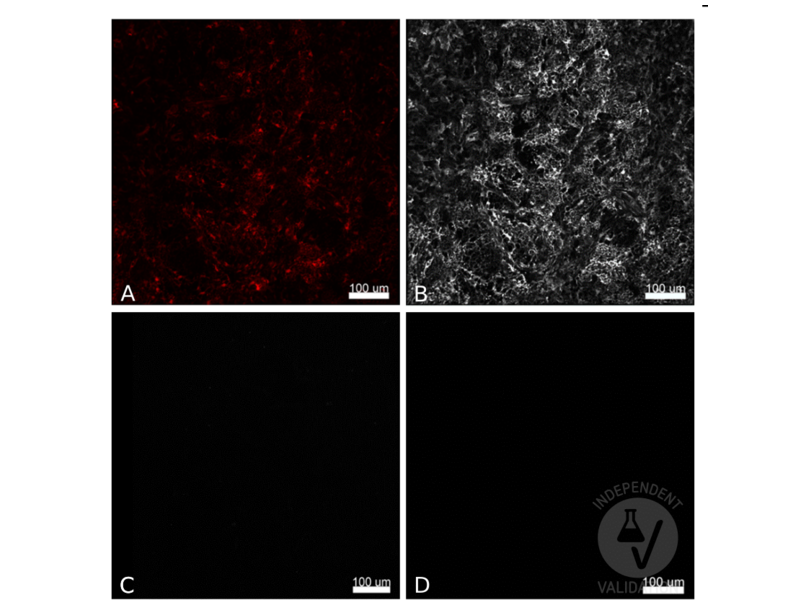
- Contrôle positif
- CX3CL1mcherry+ murine thymus
- Contrôle négative
- CX3CL1 WT murine thymus
- Conclusion
Passed. The RFP antibody ABIN129578 specifically labels RFP+ cells in CX3CL1mcherry+ murine thymus in immunofluorescence, consistent with the expression pattern of CX3CL1mcherry.
- Anticorps primaire
- ABIN1043867
- Anticorps secondaire
- donkey Fab'2 anti-rabbit A647 conjugated antibody (Jackson Immunoresearch, 711-606-152, lot 128806)
- Full Protocol
- Harvest thymus from 7 weeks old mice in 1X Dulbecco's phosphate buffered saline (DPBS) (Gibco Life Technologies, 14200-067).
- Fix mouse thymus in antigen fix (Diapath, P0014) for 2h at 4?C.
- Wash tissue in 0.1M pH7.4 phosphate buffer at for 1h at 4?C.
- Dehydrate tissue in 30% sucrose solution at 4?C ON.
- Snap freeze tissue in Tissue Freezing Medium (ElectroMicroscopy Science, 72592-C) at -80?C.
- Cut blocks into 25?m sections using a cryostat (Leica, CM3050 S).
- Transfer sections to a slide.
- Create a hydrophobic barrier on the slide around sections with Dako pen (Dako, S2002, lot 00081640).
- Place slide in a humidified chamber and rehydrate sections in 0.1 M TrisHCl pH7.4 for 10min at RT.
- Gently remove buffer by tapping slide.
- Permeabilize tissue in 0.1M TrisHCl pH7.4 containing 2% Triton X-100 (Sigma, lot 015K0039) and 0.5% BSA, for 20min at RT.
- Incubate sections with primary RFP antibody (Red Fluorescent Protein) (AA 234) (antibodies-online, ABIN129578, lot 34945) diluted 1:1000 0.1M TrisHCl pH7.4 containing 2% Triton X-100 (Sigma, batch 015K0039) and 0.5% BSA for 2h at RT.
- Incubate a no primary antibody negative control in parallel in 0.1M TrisHCl pH7.4 containing 2% Triton X-100 (Sigma, batch 015K0039) and 0.5% BSA.
- Wash slides 1x 5min in 0.1M TrisHCl pH7.4.
- Incubate sections with secondary donkey Fab'2 anti-rabbit AF647 conjugated antibody (Jackson Immunoresearch, 711-606-152, lot 128806) diluted 1:300 in 0.1M TrisHCl pH7.4 containing 2% Triton X-100 (Sigma, batch 015K0039) and 0.5% BSA for 2h at RT.
- Wash slides 1x 5min in 0.1M TrisHCl pH7.4.
- Add approximately 10µL of Slowfade Gold antifade reagent (ThermoFisher Scientific, S36937, lot 1226836) for each section and mount cover slip.
- Image acquisition on an LSM 880 (Zeiss), 20x magnification, 1000 resolution.
- Notes
Validation #101103 (Immunofluorescence)
 Validation Images
![CX3CL1 positive cells in CX3CL1mcherry murine thymus (A, mCherry) were stained with ABIN1043867 and an AF647 conjugated secondary antibody (B) or with the secondary antibody alone (C). CX3CL1 positive cells in CX3CL1 wt murine thymus were stained with ABIN1043867 and the AF647 conjugated antibody (D). Scale bar 100?m, 20x magnification, 1000 resolution]() CX3CL1 positive cells in CX3CL1mcherry murine thymus (A, mCherry) were stained with ABIN1043867 and an AF647 conjugated secondary antibody (B) or with the secondary antibody alone (C). CX3CL1 positive cells in CX3CL1 wt murine thymus were stained with ABIN1043867 and the AF647 conjugated antibody (D). Scale bar 100?m, 20x magnification, 1000 resolution
Protocole
CX3CL1 positive cells in CX3CL1mcherry murine thymus (A, mCherry) were stained with ABIN1043867 and an AF647 conjugated secondary antibody (B) or with the secondary antibody alone (C). CX3CL1 positive cells in CX3CL1 wt murine thymus were stained with ABIN1043867 and the AF647 conjugated antibody (D). Scale bar 100?m, 20x magnification, 1000 resolution
Protocole -
-
Format
- Liquid
-
Concentration
- 1.03 mg/mL
-
Buffer
- 0.02 M Potassium Phosphate, 0.15 M Sodium Chloride, pH 7.2, 0.01% (w/v) Sodium Azide
-
Agent conservateur
- Sodium azide
-
Précaution d'utilisation
- This product contains sodium azide: a POISONOUS AND HAZARDOUS SUBSTANCE which should be handled by trained staff only.
-
Conseil sur la manipulation
- Avoid cycles of freezing and thawing.
-
Stock
- 4 °C/-20 °C
-
Stockage commentaire
- Store vial at -20° C prior to opening. Aliquot contents and freeze at -20° C or below for extended storage. Centrifuge product if not completely clear after standing at room temperature. This product is stable for several weeks at 4° C as an undiluted liquid. Dilute only prior to immediate use.
-
Date de péremption
- 12 months
-
-
-
: "Epidermal Stem Cells Control Periderm Injury Repair via Matrix-Driven Specialization of Intercellular Junctions." dans: bioRxiv : the preprint server for biology, (2025) (PubMed).
: "Epidermal stem cells control periderm injury repair via matrix-driven specialization of intercellular junctions." dans: Nature communications, Vol. 16, Issue 1, pp. 8967, (2025) (PubMed).
: "Axonal injury is a targetable driver of glioblastoma progression." dans: Nature, Vol. 646, Issue 8084, pp. 452-461, (2025) (PubMed).
: "Akt is a mediator of artery specification during zebrafish development." dans: Development (Cambridge, England), Vol. 151, Issue 17, (2024) (PubMed).
: "Latrophilin-2 mediates fluid shear stress mechanotransduction at endothelial junctions." dans: The EMBO journal, Vol. 43, Issue 15, pp. 3175-3191, (2024) (PubMed).
: "Haematopoietic stem and progenitor cell heterogeneity is inherited from the embryonic endothelium." dans: Nature cell biology, Vol. 25, Issue 8, pp. 1135-1145, (2023) (PubMed).
: "Combining long-term circuit mapping and network transcriptomics with SiR-N2c." dans: Nature methods, Vol. 20, Issue 4, pp. 580-589, (2023) (PubMed).
: "Injury primes mutation-bearing astrocytes for dedifferentiation in later life." dans: Current biology : CB, (2023) (PubMed).
: "Diet suppresses glioblastoma initiation in mice by maintaining quiescence of mutation-bearing neural stem cells." dans: Developmental cell, (2023) (PubMed).
: "Molecular sensing of mechano- and ligand-dependent adhesion GPCR dissociation." dans: Nature, Vol. 615, Issue 7954, pp. 945-953, (2023) (PubMed).
: "Overexpression of Lin28A in neural progenitor cells in vivo does not lead to brain tumor formation but results in reduced spine density." dans: Acta neuropathologica communications, Vol. 9, Issue 1, pp. 185, (2022) (PubMed).
: "A versatile viral toolkit for functional discovery in the nervous system. ..." dans: Cell reports methods, Vol. 2, Issue 6, pp. 100225, (2022) (PubMed).
: "Axon guidance at the spinal cord midline-A live imaging perspective." dans: The Journal of comparative neurology, (2021) (PubMed).
: "Extrinsic Regulators of mRNA Translation in Developing Brain: Story of WNTs." dans: Cells, Vol. 10, Issue 2, (2021) (PubMed).
: "Co-activation of Sonic hedgehog and Wnt signaling in murine retinal precursor cells drives ocular lesions with features of intraocular medulloepithelioma." dans: Oncogenesis, Vol. 10, Issue 11, pp. 78, (2021) (PubMed).
: "The white matter is a pro-differentiative niche for glioblastoma." dans: Nature communications, Vol. 12, Issue 1, pp. 2184, (2021) (PubMed).
: "Antinociceptive modulation by the adhesion GPCR CIRL promotes mechanosensory signal discrimination." dans: eLife, Vol. 9, (2021) (PubMed).
: "Reticular Fibroblasts Expressing the Transcription Factor WT1 Define a Stromal Niche that Maintains and Replenishes Splenic Red Pulp Macrophages." dans: Immunity, Vol. 53, Issue 1, pp. 127-142.e7, (2020) (PubMed).
: "Transcriptional activities of human elongation factor-1α and cytomegalovirus promoter in transgenic dogs generated by somatic cell nuclear transfer." dans: PLoS ONE, Vol. 15, Issue 6, pp. e0233784, (2020) (PubMed).
: "Gastric squamous-columnar junction contains a large pool of cancer-prone immature osteopontin responsive Lgr5-CD44+ cells." dans: Nature communications, Vol. 11, Issue 1, pp. 84, (2020) (PubMed).
-
-
- RFP (Red Fluorescent Protein (RFP))
-
Autre désignation
- RFP
-
Sujet
-
Synonyms: DsRed, rDsRed, Discosoma sp. Red Fluorescent Protein, Red fluorescent protein drFP583
Fluorescent proteins such as Discosoma Red Fluorescent Protein (DsRed) from sea anemone Discosoma sp. mushroom or green fluorescent protein (GFP) from Aequorea victoria jellyfish are widely used in research practice. Fusion RFP and GFP commonly serve as marker for gene expression and protein localization. As DsRed and GFP share only 19% identity, therefore, in general, anti-GFP antibodies do not recognize DsRed protein and vice versa. Structurally, Discosoma red fluorescent protein is similar to Aequorea green fluorescent protein in terms of its overall fold (a β-can) and chromophore-formation chemistry. However, Discosoma red fluorescent protein undergoes an additional step in the chromophore maturation and obligates tetrameric structure.
Antigène
-


