HA-Tag anticorps (C-Term, N-Term)


 (1 reference)
 (2 validations)
Aperçu rapide pour HA-Tag anticorps (C-Term, N-Term) (ABIN2443910)
Antigène
Hôte
Clonalité
Conjugué
Application
Clone
-
-
Épitope
- C-Term, N-Term
-
Fonction
- HA Tag Mouse Monoclonal Antibody (HA.C5)
-
Séquence
- YPYDVPDYA
-
Specificité
- The antibody recognizes the HA epitope (YPYDVPDYA) on N-terminal, C-terminal or internal HA-tagged fusion proteins.
-
Purification
- Protein G purified
-
Immunogène
- The antibody was raised against YPYDVPDYA (HA) synthetic peptide conjugated to KLH.
-
Isotype
- IgG3
-
-
anti-HA-Tag antibody
WB, ELISA, IP, IF, FACS Hôte: Souris Monoclonal 18B11H6 unconjugated
anti-HA-Tag antibodyWB, IF, ChIP Hôte: Lapin Polyclonal RB0411 unconjugated
anti-HA-Tag antibodyWB, ELISA, IP Hôte: Lapin Polyclonal unconjugated
anti-HA-Tag antibodyReactivité: Tag WB, ELISA, IP, IF, ChIP Hôte: Lapin Polyclonal unconjugated
anti-HA-Tag (AA 98-106) antibodyAnimal-Free Reactivité: Tag WB, ELISA, IF Hôte: Lapin Monoclonal AB158-1-D11 unconjugated Recombinant Antibody
anti-HA-Tag antibodyReactivité: Humain WB Hôte: Souris Monoclonal 12CA5 unconjugated
anti-HA-Tag antibody (HRP)WB, ELISA, IP Hôte: Souris Monoclonal 5E11D8 HRP
anti-HA-Tag (AA 98-106) antibodyReactivité: Différentes espèces WB, ELISA, IF, FACS, IHC Hôte: Souris Monoclonal 16-43 unconjugated
anti-HA-Tag (AA 98-106) antibodyAnimal-Free Reactivité: Tag WB, ELISA, IF Hôte: Souris Monoclonal AB158-1-D11 unconjugated Recombinant Antibody
anti-HA-Tag antibodyReactivité: Tag WB, ELISA Hôte: Lapin Polyclonal unconjugated
-
-
Indications d'application
- WB 1:1000,IHC 1:500
-
Restrictions
- For Research Use only
-
-
- by
- Georg-August-University of Göttingen, Johann-Friedrich-Blumenbach-Institute for Zoology and Anthropology, Developmental Biology
- No.
- #100058
- Date
- 16.09.2016
- Antigène
- HA-Tag
- Numéro du lot
- 16631508212-P
- Application validée
- Western Blotting
- Contrôle positif
- HA-fusion proteins expressed in NIH/3T3 cells
- Contrôle négative
- Conclusion
- Passed, regarding sensitivity and specificity. The HA-tag antibody ABIN2443910 does specifically recognize HA-tagged proteins in whole cell lysates from human tissue culture cells with no obvious background staining.
- Anticorps primaire
- ABIN2443910
- Anticorps secondaire
- Rabbit anti-Mouse IgG (whole molecule), HRP-linked (Sigma-Aldrich, A9044, lot number: 034M4761)
- Full Protocol
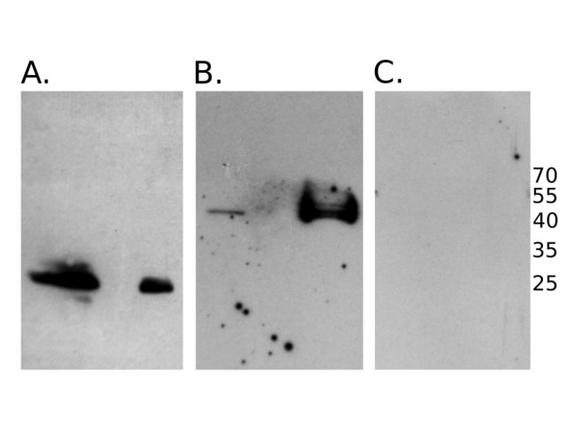
- Cultured NIH/3T3 cells heterologously expressing either a 26kDa (Fig. A) or 43kDa (Fig. B) HA-tag-fusion protein were rinsed with PBS and lysed in 6M Urea/20mM Tris.
- 40µg total protein were used for Western blot analysis.
- Proteins were denatured and separated on 10% SDS-PAGE (Laemmli 1970) and blotted to Amersham Protran Premium 0.2 NC (GE Healthcare, 10600004, lot A10043108) (Towbin et al., 1979).
- Blocking of membrane in 5% skim milk in TBST (50 mM Tris-HCl, pH 7.4, 150mM NaCl, 0.1% Tween 20) for 30min at RT.
- Incubation with primary antibody ABIN2443910 diluted 1:600 in 5% skim milk in TBST overnight at 4°C.
- Washing in TBST for 30min at RT.
- Incubation with secondary antibody secondary antibody: rabbit-anti-mouse IgG (whole molecule), HRP-linked (Sigma-Aldrich, A9044, lot 034M4761) diluted 1:10000 in 5% skim milk in TBST for 45min at RT.
- Washing in TBST for 30-45min at RT.
- Chemiluminescence detection using Clarity Western ECL Substrate (BioRad, 170-5061) according to the supplier’s recommendations and image capture via X-ray films.
- Notes
- A dilution factor <1000 is recommended for Western blot on cellular lysates with ABIN2443910.
Validation #100058 (Western Blotting)
 Validation Images
![HA-tagged fusion proteins were expressed in NIH/3T3 cells. Whole cell lysates were separated by SDS-PAGE and the tagged protein detected using anti HA-tag antibody ABIN2443910 diluted 1:600. Expected molecular mass of the fusion proteins were approximately 26kDa (A.) and 43kDa (B.) respectively. The left lane corresponds in both blots A. and B. to the pellet, the right to the supernatant after centrifugation. Exposition time for image capture on X-ray films: 1min (A.) and 30min (B.). For C., samples were prepared as for B. but the anti HA-tag antibody ABIN2443910 was diluted 1:1000. Exposure time: 15min.]() HA-tagged fusion proteins were expressed in NIH/3T3 cells. Whole cell lysates were separated by SDS-PAGE and the tagged protein detected using anti HA-tag antibody ABIN2443910 diluted 1:600. Expected molecular mass of the fusion proteins were approximately 26kDa (A.) and 43kDa (B.) respectively. The left lane corresponds in both blots A. and B. to the pellet, the right to the supernatant after centrifugation. Exposition time for image capture on X-ray films: 1min (A.) and 30min (B.). For C., samples were prepared as for B. but the anti HA-tag antibody ABIN2443910 was diluted 1:1000. Exposure time: 15min.
Protocole
HA-tagged fusion proteins were expressed in NIH/3T3 cells. Whole cell lysates were separated by SDS-PAGE and the tagged protein detected using anti HA-tag antibody ABIN2443910 diluted 1:600. Expected molecular mass of the fusion proteins were approximately 26kDa (A.) and 43kDa (B.) respectively. The left lane corresponds in both blots A. and B. to the pellet, the right to the supernatant after centrifugation. Exposition time for image capture on X-ray films: 1min (A.) and 30min (B.). For C., samples were prepared as for B. but the anti HA-tag antibody ABIN2443910 was diluted 1:1000. Exposure time: 15min.
Protocole -
- by
- Georg-August-University of Göttingen, Johann-Friedrich-Blumenbach-Institute for Zoology and Anthropology, Developmental Biology
- No.
- #100059
- Date
- 16.09.2016
- Antigène
- HA-Tag
- Numéro du lot
- 16631508212-P
- Application validée
- Immunocytochemistry
- Contrôle positif
- HA-fusion proteins transiently expressed in NIH/3T3 cells
- Contrôle négative
- Conclusion
- Passed, regarding sensitivity and specificity. The antibody specifically detects the HA-fusion protein in immuno-cytochemical experiments. No obvious background staining was found.
- Anticorps primaire
- ABIN2443910
- Anticorps secondaire
- MFP488 goat anti-mouse IgG (H+L) (MoBiTec, MFP-A1029, lot 2803101)
- Full Protocol
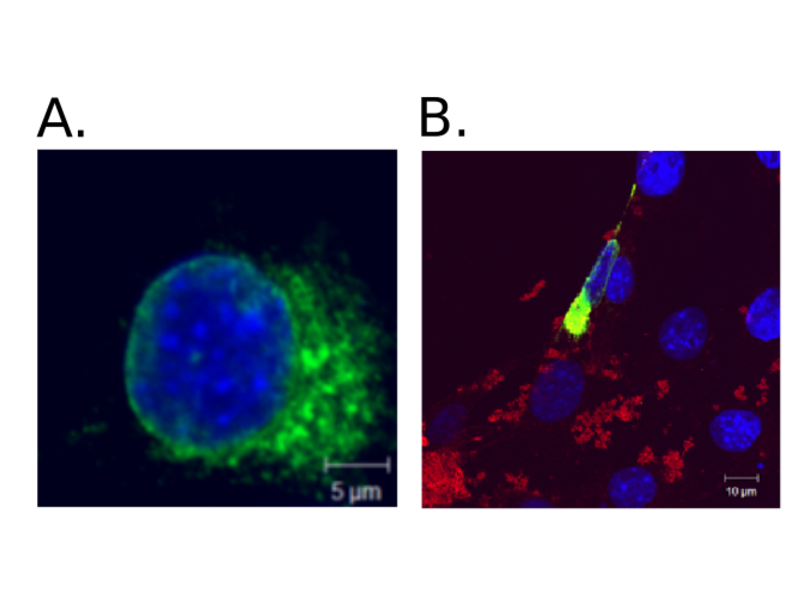
- NIH/3T3 cells (ATCC) were grown on cover slips in DMEM, 10% fetal bovine serum, 5% penicillin/streptomycin (all Gibco) at 37°C in 5% CO2.
- Cells were transfected using Metafectene Pro (Biontex, T040-1.0, lot AD1.13) according to the manual with a plasmid encoding an HA-tag fusion protein that is expected to localize to the nuclear membrane and the cytoplasm.
- 24h post-transfection cells were fixed in 3.7% paraformaldehyde in PBS for 15min and processed for immuno-cytology.
- Unspecific binding sites were blocked in PBST (phosphate buffered saline containing 0.15% bovine serum albumin, 0.1% Tween-20) for 1h.
- Cells were incubated with the primary antibody ABIN2443910 diluted 1:100 in PBS at 4°C overnight.
- Cells were washed in TBST (50mM Tris-HCl, pH 7.4, 150mM NaCl, 0.1% Tween 20) for 15min.
- Incubation with the secondary antibody MFP488 goat anti-mouse IgG (H+L) (MoBiTec, MFP-A1029, lot 2803101) diluted 1:600 in PBS and DAPI (stock solution 10µg/µl, diluted 1:500) for 1h at 37°C.
- Cells were washed in TBST for 15min.
- Cells were embedded in Fluorescent Mounting Medium (Dako, S3023, lot 10090890).
- Images were taken by confocal microscopy (LSM 510, Zeiss), and processed using Adobe Photoshop 5.0.
- Notes
- none
Validation #100059 (Immunocytochemistry)
 Validation Images
![HA-tagged fusion proteins were expressed in NIH/3T3 cells. The protein of interest is expected to localize to the nuclear membrane and the cytoplasm. A. The fusion protein was revealed via its HA-tag at the nuclear membrane and in the cytoplasm using ABIN2443910 (green; DAPI counterstain in blue). B. Co-staining of the fusion protein with using using anti-HA tag antibody ABIN2443910 (green) and an anti-myc-tag antibody (red; unspecific background staining caused by the transfection reagent). DAPI staining was used to visualize the nucleus (blue).]() HA-tagged fusion proteins were expressed in NIH/3T3 cells. The protein of interest is expected to localize to the nuclear membrane and the cytoplasm. A. The fusion protein was revealed via its HA-tag at the nuclear membrane and in the cytoplasm using ABIN2443910 (green; DAPI counterstain in blue). B. Co-staining of the fusion protein with using using anti-HA tag antibody ABIN2443910 (green) and an anti-myc-tag antibody (red; unspecific background staining caused by the transfection reagent). DAPI staining was used to visualize the nucleus (blue).
Protocole
HA-tagged fusion proteins were expressed in NIH/3T3 cells. The protein of interest is expected to localize to the nuclear membrane and the cytoplasm. A. The fusion protein was revealed via its HA-tag at the nuclear membrane and in the cytoplasm using ABIN2443910 (green; DAPI counterstain in blue). B. Co-staining of the fusion protein with using using anti-HA tag antibody ABIN2443910 (green) and an anti-myc-tag antibody (red; unspecific background staining caused by the transfection reagent). DAPI staining was used to visualize the nucleus (blue).
Protocole -
-
Format
- Lyophilized
-
Reconstitution
- If reconstituted with deionized water in 100 μL: WB 1:1000-3,000, IHC 1:500-2,000. Optimal dilution has to be determined by the user.
-
Buffer
- Lyophilized protein G purified in PBS pH 7.4
-
Agent conservateur
- Without preservative
-
Stock
- 4 °C,-20 °C,-80 °C
-
Stockage commentaire
- Lyophilized antibodies can be kept at 4°C for up to 3 months and should be kept at -20°C for long-term storage (2 years). To avoid freeze-thaw cycles, reconstituted antibodies should be aliquoted before freezing for long-term (1 year) storage (-80°C) or kept at 4°C for short-term usage (2 months). For maximum recovery of product, centrifuge the original vial prior to removing the cap. Further dilutions can be made with the assay buffer. After the maximum long-term storage period (2 years lyophilized or 1 year reconstituted) antibodies should be tested in your assay with a standard sample to verify if you have noticed any decrease in their efficacy. To limit antibody loss or degradation, BSA (final concentration 1%) and sodium azide (final concentration 0.02%) can be added to the suggested first dilution. It is important to first verify if those preservatives are compatible with your assay.
-
Date de péremption
- 24 months
-
-
-
: "APOBEC3-mediated restriction of RNA virus replication." dans: Scientific reports, Vol. 8, Issue 1, pp. 5960, (2019) (PubMed).
-
-
- HA-Tag
-
Autre désignation
- HA Tag
-
Classe de substances
- Tag
-
Sujet
- Epitope tags have application in the labeling, isolation and detection of proteins using immunoblotting, Immunoprecipitation and immunostaining techniques. Epitope tags can be used, by affinity chromatography, to separate recombinant, overexpressed protein from wild-type protein expressed by the host organism. Due its small size the protein's biochemical properties seem not to be affected by the tag protein. Human influenza hemagglutinin (HA) is a surface glycoprotein required for the infectivity of human by the virus. The HA tag corresponds to the amino acids 98-106 (YPYDVPDYA) of the HA protein. It has been extensively used as a general epitope tag in expression vectors. Many recombinant proteins have been engineered to express the HA tag.
Antigène
-