Histone 3 anticorps (H3K36me3)
 (4 références)
 (1 validation)
Aperçu rapide pour Histone 3 anticorps (H3K36me3) (ABIN2668403)
Antigène
Voir toutes Histone 3 (H3) AnticorpsReactivité
Hôte
Clonalité
Conjugué
Application
Clone
-
-
Épitope
- H3K36me3
-
Fonction
- Histone H3K36me3 antibody (mAb) (Clone MABI 0333)
-
Purification
- Protein G Chromatography
-
Immunogène
- This Histone H3 trimethyl Lys36 antibody was raised against a peptide containing trimethyl Lys36 of human Histone H3.
-
Isotype
- IgG1
-
-
anti-Histone H3 (H3) (3meLys4) antibody
H3 Reactivité: Humain, Saccharomyces cerevisiae WB, IF, ChIP, ICC, DB, ChIP-seq, CUT&Tag, CUT&RUN Hôte: Lapin Polyclonal unconjugated
anti-Histone H3 (H3) (H3K4me) antibodyH3 Reactivité: Humain WB, IF, ChIP, IP, ChIP-seq, CUT&RUN Hôte: Lapin Polyclonal unconjugated
anti-Histone H3 (H3) (H3K27ac) antibodyH3 Reactivité: Humain, Saccharomyces cerevisiae WB, IF, IHC, ChIP, ICC, DB, ChIP-seq, CUT&Tag Hôte: Lapin Polyclonal unconjugated
anti-Histone H3 (H3) (H3K9ac) antibodyH3 Reactivité: Humain, Souris WB, IF, ChIP, ICC, DB, ChIP-seq, CUT&Tag Hôte: Lapin Polyclonal unconjugated
anti-Histone H3 (H3) (H3K27me) antibodyH3 Reactivité: Humain WB, IF, IHC, ChIP, IP, ChIP-seq Hôte: Lapin Polyclonal unconjugated
anti-Histone H3 (H3) (AA 71-136) antibodyH3 Reactivité: Humain WB, FACS, IHC (p), IF (cc), IF (p), IHC (fro) Hôte: Lapin Polyclonal unconjugated
anti-Histone H3 (H3) (H3R17me) antibodyH3 Reactivité: Humain WB, IF, IP Hôte: Lapin Polyclonal unconjugated
anti-Histone H3 (H3) (H3K36me3) antibodyH3 Reactivité: Humain WB, IF, IHC, ChIP, IP, ChIP-seq Hôte: Lapin Polyclonal unconjugated
anti-Histone H3 (H3) (H3K36me2) antibodyH3 Reactivité: Humain WB, IF, IHC, ChIP, IP, ChIP-seq Hôte: Lapin Polyclonal unconjugated
anti-Histone H3 (H3) (H3R2me2) antibodyH3 Reactivité: Humain WB, IF, ChIP, IP, ChIP-seq Hôte: Lapin Polyclonal unconjugated
-
-
Indications d'application
- ChIP: 5 - 10 μg per ChIP ChIP-Seq: 5 - 10 μg each WB: 0.5 - 2 μg/mL dilution DB: 0.5 - 2 μg/mL dilution ChIP-Seq validation was performed by Active Motif's Epigenetics Services, the complete data set is available in the UCSC Genome Browser by clicking here.
-
Restrictions
- For Research Use only
-
-
- by
- Anna Nordin and Claudio Cantù; Cantù Lab, Gene Regulation during Development and Disease, Linköping University
- No.
- #104510
- Date
- 14.08.2023
- Antigène
- H3K36me3
- Numéro du lot
- 20822015
- Application validée
- Cleavage Under Targets and Release Using Nuclease
- Contrôle positif
Polyclonal rabbit anti-H3K4me (antibodies-online, ABIN3023251)
- Contrôle négative
Polyclonal guinea pig anti-rabbit IgG (antibodies-online, ABIN101961)
- Conclusion
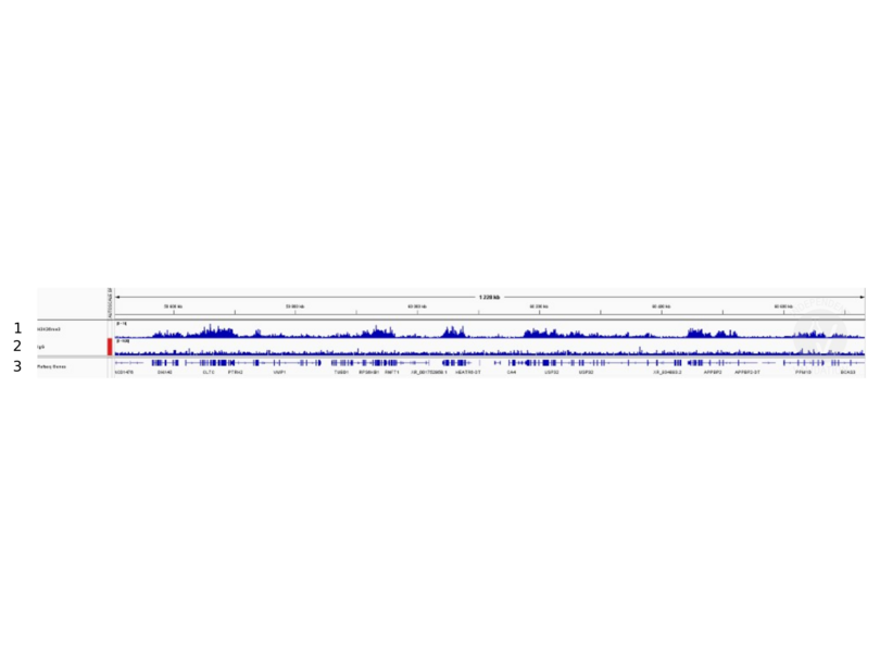
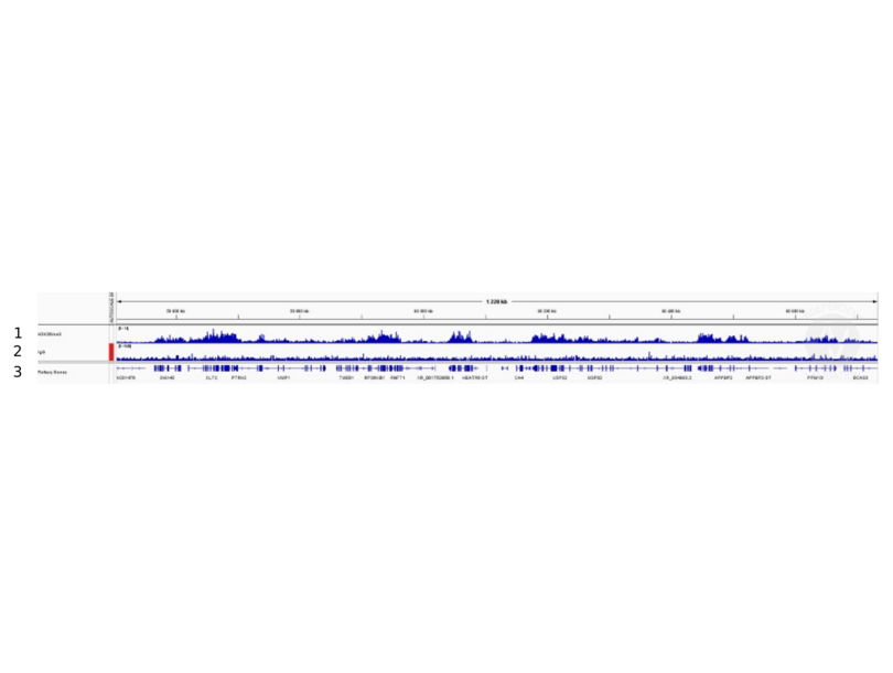
Passed. ABIN2668403 allows for specific targeting of H3K36me3 in human cells using CUT&RUN.
- Anticorps primaire
- ABIN2668403
- Anticorps secondaire
- Full Protocol
- Cell harvest
- Harvest 50,000 human fibroblast cells per antibody.
- Centrifuge cell solution 3 min at 600 x g at RT.
- Remove the liquid carefully.
- Gently resuspend cells in 1 mL of Nuclear Extraction Buffer (20 mM HEPES-KOH pH 8.2, 20% Glycerol, 0,05% IGEPAL, 0.5 mM Spermidine, 10 mM KCl, Roche Complete Protease Inhibitor EDTA-free).
- Move the solution to a 2 mL centrifuge tube.
- Pellet the nuclei 800 x g for 5 min.
- Repeat the NE Buffer wash twice for a total of three washes.
- Resuspend the nuclei in 20 µL NE Buffer per sample.
- Concanavalin A beads preparation
- Prepare one 2 mL microcentrifuge tube.
- Gently resuspend the magnetic Concanavalin A Beads (antibodies-online, ABIN6923139).
- Pipette 10 µL Con A Beads slurry for each sample into the 1.5 mL microcentrifuge tube.
- Place the tube on a magnet stand until the fluid is clear. Remove the liquid carefully.
- Remove the microcentrifuge tube from the magnetic stand.
- Pipette 1 mL Binding Buffer (20 mM HEPES pH 7.5, 10 mM KCl, 1 mM CaCl2, 1 mM MnCl2) into each tube and resuspend ConA beads by gentle pipetting.
- Spin down the liquid from the lid with a quick pulse in a table-top centrifuge.
- Place the tubes on a magnet stand until the fluid is clear. Remove the liquid carefully.
- Remove the microcentrifuge tube from the magnetic stand.
- Repeat twice for a total of three washes.
- Gently resuspend the ConA Beads in a volume of Binding Buffer corresponding to the original volume of bead slurry, i.e. 10 µL per sample.
- Nuclei immobilization – binding to Concanavalin A beads
- Carefully vortex the nuclei suspension and add 10 µL of the Con A beads in Binding Buffer to the cell suspension for each sample.
- Close tube tightly incubates 10 min at 4 °C.
- Put the 2 mL tube on the magnet stand and when the liquid is clear remove the supernatant.
- Resuspend the beads in 1 mL of EDTA wash buffer (20 mM HEPES pH 7.5, 150 mM NaCl, 0.5 mM Spermidine, Roche Complete Protease Inhibitor EDTA-free, 2mM EDTA).
- Incubate 5 min at RT.
- Place the tube on the magnet stand and when the liquid is clear remove the supernatant.
- Resuspend the beads in 200µl of Wash Buffer (20 mM HEPES pH 7.5, 150 mM NaCl, 0.5 mM Spermidine, Roche Complete Protease Inhibitor EDTA-free) for each sample.
- Cell permeabilization and primary antibody binding
- Divide nuclei suspension into separate PCR tubes, one for each antibody (200 µL per sample).
- Add 2 µL antibody (anti-H3K36me3 antibody ABIN2668403, anti-H3K4me positive control antibody ABIN3023251, and guinea pig anti-rabbit IgG negative control antibody ABIN101961) to the respective tube, corresponding to a 1:100 dilution.
- Incubate ON at 4 °C.
- Place the tubes on a magnet stand until the fluid is clear. Remove the liquid carefully.
- Remove the microcentrifuge tubes from the magnetic stand.
- Wash with 200 µL of Wash Buffer using a multichannel pipette to accelerate the process.
- Repeat the wash five times for a total of six washes.
- pAG-MNase Binding
- Prepare a 1.5 mL microcentrifuge tube containing 200 µL of pAG mix for each sample (200 µl of wash buffer + 120 ng pAG-MNase per sample).
- Place the PCR tubes with the sample on a magnet stand until the fluid is clear. Remove the liquid carefully.
- Remove tubes from the magnetic stand.
- Resuspend the beads in 200 µL of pAG-MNase premix.
- Incubate for 30 min at 4 °C.
- Place the tubes on a magnet stand until the fluid is clear. Remove the liquid carefully.
- Remove the microcentrifuge tubes from the magnetic stand.
- Wash with 200 µL of Wash Buffer using a multichannel pipette to accelerate the process.
- Repeat the wash for a total of five washes.
- Resuspend in 200 µL of Wash Buffer.
- MNase digestion and release of pAG-MNase-antibody-chromatin complexes
- Place PCR tubes on ice and allow to chill.
- Prepare a 1.5 mL microcentrifuge tube with 51 µl of 2 mM CaCl2 mix per sample (50 µl Wash Buffer + 1 µL 100 mM CaCl2) and let it chill on ice.
- Always in ice, place the samples on the magnetic rack and when the liquid is clear remove the supernatant.
- Resuspend the samples in 50 µl of the 2 mM CaCl2 mix and incubate in ice for exactly 30 min.
- Place the sample on the magnet stand and when the liquid is clear move the supernatant in fresh collection tubes with 3µl of EDTA/EGTA 0.25M (Digestion buffer).
- Resuspend the sample in 47 µl of 1x Urea STOP Buffer (8.5 M Urea, 100 mM NaCl, 2 mM EGTA, 2 mM EDTA, 0,5% IGEPAL).
- Incubate the samples 1 h at 4 °C.
- Transfer the supernatant containing the pAG-MNase-bound digested chromatin fragments to the previously collected digestion buffer.
- DNA Clean up
- Take the Mag-Bind® TotalPure NGS beads (Omega Bio-Tek, M1378-01) from the storage and wait until they are at RT.
- Add 2x volume of beads to each sample (e.g. 100 µL of beads for 50 µL of sample).
- Incubate the beads and the sample for 15 min at RT.
- During incubation prepare fresh EtOH 80%.
- Place the PCR tubes on a magnet stand and when the liquid is clear remove the supernatant.
- Add 200 µl of fresh 80% EtOH to the sample without disturbing the beads (Important!!! Do NOT resuspend the beads or remove the tubes from the magnet stand or the sample will be lost).
- Incubate 30 sec at RT.
- Remove the EtOH from the sample.
- Repeat the wash with 80% EtOH.
- Resuspend the beads in 25 µL of 10 mM Tris.
- Incubate the sample for 2 min at RT.
- Repeat the 2x beads clean up as described before (this time with 50 µL of beads for each sample).
- Resuspend the beads + DNA in 20 µL of 10 mM Tris.
- Library preparation and sequencing
- Prepare libraries using KAPA HyperPrep Kit using KAPA Dual-Indexed adapters according to protocol.
- Sequence samples on an Illumina NextSeq 500 sequencer, using a NextSeq 500/550 High Output Kit v2.5 (75 Cycles), 36bp PE.
- Bioinformatics
- Align reads the human genome (hg38) using bowtie78 with settings -X 700 -m1 -v 3. Remove duplicate reads, and sort files using samtools. Filter mapped reads for size, keeping only reads with a fragment size at or below 120 base pairs.
- Generate bedgraph files using bedtools genomecov.
- Call peaks using SEACR version 1.3, in relaxed mode, normalizing to the negative control.
- Notes
Validation #104510 (Cleavage Under Targets and Release Using Nuclease)
 Validation Images
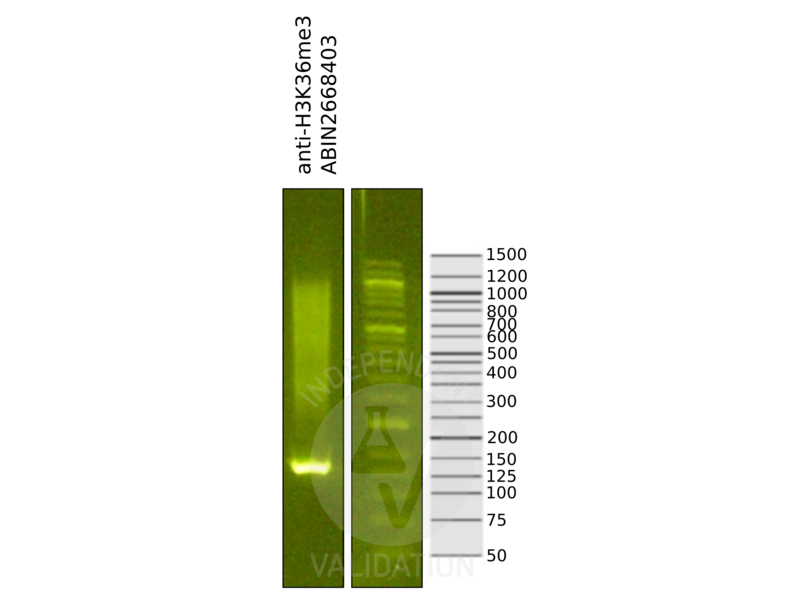
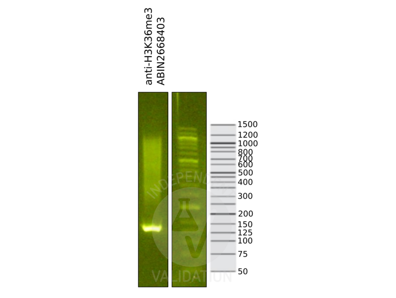
![Library profiles comparing fragment size distributions on an E-Gel EX 2% agarose gel (Thermo Fisher). Fragments obtained from CUT&RUN using anti-H3K36me3 antibody ABIN2668403 (left) after library preparation, compared to the E-Gel Sizing DNA Ladder (Thermo Fisher) (right).]() Library profiles comparing fragment size distributions on an E-Gel EX 2% agarose gel (Thermo Fisher). Fragments obtained from CUT&RUN using anti-H3K36me3 antibody ABIN2668403 (left) after library preparation, compared to the E-Gel Sizing DNA Ladder (Thermo Fisher) (right).
Library profiles comparing fragment size distributions on an E-Gel EX 2% agarose gel (Thermo Fisher). Fragments obtained from CUT&RUN using anti-H3K36me3 antibody ABIN2668403 (left) after library preparation, compared to the E-Gel Sizing DNA Ladder (Thermo Fisher) (right).
![1. Alignment tracks from CUT&RUN targeting H3K36me3 in human fibroblast cells using antibody ABIN2668403 showing the USP32 locus. 2. Alignment tracks for CUT&RUN with the IgG negative control ABIN101961. 3. RefSeq Genes.]() 1. Alignment tracks from CUT&RUN targeting H3K36me3 in human fibroblast cells using antibody ABIN2668403 showing the USP32 locus. 2. Alignment tracks for CUT&RUN with the IgG negative control ABIN101961. 3. RefSeq Genes.
Protocole
1. Alignment tracks from CUT&RUN targeting H3K36me3 in human fibroblast cells using antibody ABIN2668403 showing the USP32 locus. 2. Alignment tracks for CUT&RUN with the IgG negative control ABIN101961. 3. RefSeq Genes.
Protocole -
-
Concentration
- 0.44 μg/μL
-
Buffer
- PBS pH 7.5 containing 30 % glycerol, 0.3 M NaCl, and 0.035 % sodium azide.
-
Agent conservateur
- Sodium azide
-
Précaution d'utilisation
- This product contains Sodium azide: a POISONOUS AND HAZARDOUS SUBSTANCE which should be handled by trained staff only.
-
Conseil sur la manipulation
- Avoid repeated freeze/thaw cycles by aliquoting items into single-use fractions,Keep all reagents on ice when not in storage
-
Stock
- -20 °C
-
Stockage commentaire
- Some products may be shipped at room temperature. This will not affect their stability or performance. Avoid repeated freeze/thaw cycles by aliquoting items into single-use fractions for storage at -20°C for up to 2 years. Keep all reagents on ice when not in storage.
-
Date de péremption
- 24 months
-
-
-
: "RSC-Associated Subnucleosomes Define MNase-Sensitive Promoters in Yeast." dans: Molecular cell, Vol. 73, Issue 2, pp. 238-249.e3, (2019) (PubMed).
: "The Meiotic Recombination Activator PRDM9 Trimethylates Both H3K36 and H3K4 at Recombination Hotspots In Vivo." dans: PLoS genetics, Vol. 12, Issue 6, pp. e1006146, (2016) (PubMed).
: "Arabidopsis MRG domain proteins bridge two histone modifications to elevate expression of flowering genes." dans: Nucleic acids research, Vol. 42, Issue 17, pp. 10960-74, (2014) (PubMed).
: "The histone H3K36 methyltransferase MES-4 acts epigenetically to transmit the memory of germline gene expression to progeny." dans: PLoS genetics, Vol. 6, Issue 9, pp. e1001091, (2010) (PubMed).
-
-
- Histone 3 (H3) (Histone H3 (H3))
-
Autre désignation
- Histone H3
-
Sujet
- Histone H3 is one of the core components of the nucleosome. The nucleosome is the smallest subunit of chromatin and consists of 147 base pairs of DNA wrapped around an octamer of core histone proteins (two each of Histone H2A, Histone H2B, Histone H3 and Histone H4). Chromatin is subject to a variety of chemical modifications, including post-translational modifications of the histone proteins and the methylation of cytosine residues in the DNA. Reported histone modifications include acetylation, methylation, phosphorylation, ubiquitylation, glycosylation, ADP-ribosylation, carbonylation and SUMOylation, these modifications play a major role in regulating gene expression. The methylation of histones can occur on two different residues: arginine or lysine. Histone methylation can be associated with transcriptional activation or repression, depending on the methylated residue. Histone H3 is methylated at lysine 36 by the Set2 (yeast) and NSD1 (mammals) methyltransferases. Dimethylation of lysine 36 of histone H3 is involved with transcriptional elongation by RNA pol II holoenzyme and is a marker of transcribed genes.
-
Poids moléculaire
- 17 kDa
-
ID gène
- 3020
-
NCBI Accession
- NP_003522
Antigène
-