SPNS2 anticorps (N-Term)
 (1 validation)
Aperçu rapide pour SPNS2 anticorps (N-Term) (ABIN2786494)
Antigène
Voir toutes SPNS2 AnticorpsReactivité
Hôte
Clonalité
Conjugué
Application
-
-
Épitope
- N-Term
-
Séquence
- PPGTPGTPGC AATAKGPGAQ QPKPASLGRG RGAAAAILSL GNVLNYLDRY
-
Homologie
- Human: 100%, Mouse: 85%, Pig: 100%, Rabbit: 92%, Rat: 93%
-
Attributs du produit
- This is a rabbit polyclonal antibody against SPNS2. It was validated on Western Blot using a cell lysate as a positive control.
-
Purification
- Affinity Purified
-
Immunogène
- The immunogen is a synthetic peptide directed towards the N terminal region of human SPNS2
-
-
anti-Spinster Homolog 2 (SPNS2) (AA 68-94), (N-Term) antibody
SPNS2 Reactivité: Humain WB, IHC (p), FACS Hôte: Lapin Polyclonal RB38003 unconjugated
anti-Spinster Homolog 2 (SPNS2) (AA 71-120) antibodySPNS2 Reactivité: Humain, Souris, Singe, Porc, Roussette (Chauve-souris) WB, IHC, IHC (p) Hôte: Lapin Polyclonal unconjugated
anti-Spinster Homolog 2 (SPNS2) (AA 1-140) antibodySPNS2 Reactivité: Humain ELISA, IF Hôte: Lapin Polyclonal unconjugated
-
-
Indications d'application
- Optimal working dilutions should be determined experimentally by the investigator.
-
Commentaires
-
Antigen size: 549 AA
-
Restrictions
- For Research Use only
-
-
- by
- Puissant lab, Institute of Hematology, Inserm U944, CNRS UMR 7212
- No.
- #101829
- Date
- 01.02.2018
- Antigène
- SPNS2
- Numéro du lot
- QC66273-42972
- Application validée
- Western Blotting
- Contrôle positif
- Baf-3 cells/ MLL-AF9 murine primary cell model
- Contrôle négative
- Baf-3 or MLL-AF9 murine primary cell model silenced for SPNS2 gene with 2 different shRNA
- Conclusion
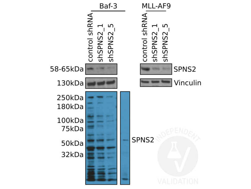
Passed. ABIN2786494 specifically recognizes SPNS2 in murine Baf-3 cells or MLL-AF9 primary cell model.
- Anticorps primaire
- ABIN2786494
- Anticorps secondaire
- mouse anti-rabbit HRP conjugated antibody (GE Healthcare, NA9340V)
- Full Protocol
- Grow Baf-3 or MLL-AF9 cells in RPMI 1640 1x (Gibco, 21875-059) supplemented with 10% FBS (Sigma-Aldrich, F6178, lot 16G148) and 1x Penicillin/Streptomycin (Gibco, 15140-122), at 37°C and 5% CO2 in 10ml on a flask to 70% confluency.
- Transduce 3x10⁶ cells with 35µl of concentrated retrovirus, spin-infection (3h at 37°C to 1200 rpm).
- Grow cells for 3d.
- Induce and select cells.
- Grow cells for 5d.
- Sort and collect cells (doxycycline).
- Lyse 7x10⁵ cells in 25µl per well in cold SDS Lysis Buffer (Cell Signaling).
- Determine total protein content of the lysates using Bradford assay (Bio-Rad, 5000006).
- Denature 20µg of total protein for 7min at 95°C in 6µl Laemmli SDS sample buffer and subsequently separate them on a denaturing NuPAGE 4-12% Bis-Tris Protein Gels (Invitrogen,NP0321BOX ) in an ELECTROPHORESIS CHAMBER for 2h at 150V.
- Transfer proteins onto PVDF membrane (Millipore) with a Western blotting system for 1h 40min at 300mA.
- Block the membrane with blocking buffer (2.75% gelatine, 30% BSA, 10mM Tris, NaCl, EDTA, tween 20) for 1h at RT.
- Incubation with rabbit anti-SPNS2 antibody (antibodies-online, ABIN2786494, lot QC66273-42972) diluted 1:500 in blocking buffer for 16h at 4°C.
- Wash membrane 3x for 10min with tween TBS buffer.
- Incubation with secondary mouse anti-rabbit HRP conjugated antibody (GE Healthcare, NA9340V) diluted 1:5000 in blocking buffer for 1h at RT.
- Wash membrane 3x for 10min with TBST buffer.
- Reveal protein bands using Pierce ECL Western Blotting Substrate (ThermoFisher Scientific, 32106) on a Li-Cor imaging machine.
- Notes
The SPNS2 antibody ABIN2786494 reveals a protein of the expected molecular weight for SPNS2 in lysates of murine Baf-3 cells or MLL-AF9 primary cell model.
ABIN2786494 does show some unspecific binding but the SPNS2 band itself is clear and only visible in the positive but not the negative controls.
Validation #101829 (Western Blotting)
 Validation Images
![Western blot analysis of SPNS2 expression in mouse Baf-3 (left) and MLL-AF9 cells (right) using ABIN2786494. Cells were transduced either with a control shRNA (control shRNA) or one of two SPNS2 shRNA (shSPNS2_1 and shSPNS2_5).]() Western blot analysis of SPNS2 expression in mouse Baf-3 (left) and MLL-AF9 cells (right) using ABIN2786494. Cells were transduced either with a control shRNA (control shRNA) or one of two SPNS2 shRNA (shSPNS2_1 and shSPNS2_5).
Protocole
Western blot analysis of SPNS2 expression in mouse Baf-3 (left) and MLL-AF9 cells (right) using ABIN2786494. Cells were transduced either with a control shRNA (control shRNA) or one of two SPNS2 shRNA (shSPNS2_1 and shSPNS2_5).
Protocole -
-
Format
- Liquid
-
Concentration
- Lot specific
-
Buffer
- Liquid. Purified antibody supplied in 1x PBS buffer with 0.09 % (w/v) sodium azide and 2 % sucrose.
-
Agent conservateur
- Sodium azide
-
Précaution d'utilisation
- This product contains Sodium azide: a POISONOUS AND HAZARDOUS SUBSTANCE which should be handled by trained staff only.
-
Conseil sur la manipulation
- Avoid repeated freeze-thaw cycles.
-
Stock
- -20 °C
-
Stockage commentaire
- For short term use, store at 2-8°C up to 1 week. For long term storage, store at -20°C in small aliquots to prevent freeze-thaw cycles.
-
-
- SPNS2 (Spinster Homolog 2 (SPNS2))
-
Autre désignation
- SPNS2
-
Sujet
-
SPNS2 is the sphingolipid transporter required for migration of myocardial precursors. SPNS2 transports sphingosine 1-phosphate (S1P), a secreted lipid mediator that plays critical roles in cardiovascular, immunological, and neural development and function. SPNS2 mediates the export of S1P from cells in the extraembryonic yolk syncytial layer (YSL), thereby regulating myocardial precursor migration.
Alias Symbols: -
Protein Size: 549 -
Poids moléculaire
- 60 kDa
-
ID gène
- 124976
-
NCBI Accession
- NM_001124758, NP_001118230
-
UniProt
- Q8IVW8
Antigène
-


