MR1 anticorps (AA 201-300)


 (3 validations)
Aperçu rapide pour MR1 anticorps (AA 201-300) (ABIN516526)
Antigène
Voir toutes MR1 AnticorpsReactivité
Hôte
Clonalité
Conjugué
Application
Clone
-
-
Épitope
- AA 201-300
-
Fonction
- Mouse monoclonal antibody raised against a partial recombinant MR1.
-
Séquence
- TEPPLVRVNR KETFPGVTAL FCKAHGFYPP EIYMTWMKNG EEIVQEIDYG DILPSGDGTY QAWASIELDP QSSNLYSCHV EHCGVHMVLQ VPQESETIPL
-
Réactivité croisée
- Humain
-
Attributs du produit
- Antibody Reactive Against Recombinant Protein.
-
Immunogène
- MR1 (NP_001522, 201 a.a. ~ 300 a.a) partial recombinant protein with GST tag. MW of the GST tag alone is 26 KDa.
-
Isotype
- IgG1
-
-
anti-Major Histocompatibility Complex, Class I-Related (MR1) (AA 312-341), (C-Term) antibody
MR1 Reactivité: Humain WB Hôte: Lapin Polyclonal RB37199 unconjugated
anti-Major Histocompatibility Complex, Class I-Related (MR1) (AA 150-253) antibodyMR1 Reactivité: Humain ELISA, WB, IHC, IF Hôte: Lapin Polyclonal unconjugated
anti-Major Histocompatibility Complex, Class I-Related (MR1) (AA 20-260) antibodyMR1 Reactivité: Humain WB, IF Hôte: Lapin Polyclonal unconjugated
anti-Major Histocompatibility Complex, Class I-Related (MR1) (AA 150-253) antibodyMR1 Reactivité: Humain, Souris ELISA, WB, IHC, IF/ICC Hôte: Lapin Polyclonal unconjugated
anti-Major Histocompatibility Complex, Class I-Related (MR1) (AA 20-260) antibodyMR1 Reactivité: Humain ELISA, IHC Hôte: Lapin Polyclonal unconjugated
anti-Major Histocompatibility Complex, Class I-Related (MR1) (AA 201-300) antibodyMR1 Reactivité: Humain ELISA, WB Hôte: Souris Polyclonal unconjugated
-
-
Indications d'application
- Optimal working dilution should be determined by the investigator.
-
Restrictions
- For Research Use only
-
-
- by
- Dr. Randy Brutkiewicz Laboratory, Department of Microbiology and Immunology, Indiana University School of Medicine
- No.
- #101752
- Date
- 20.02.2018
- Antigène
- MR1
- Numéro du lot
- 12045-5B5
- Application validée
- Immunocytochemistry
- Contrôle positif
- HEK293 cells transfected with human MR1 cDNA
- Contrôle négative
- HEK293 cells transfected with plasmid vector only
- Conclusion
Passed. The MR1 antibody ABIN516526 specifically labels the targeted antigen in HEK293 ectopically expressing human MR1 in ICC.
- Anticorps primaire
- ABIN516526
- Anticorps secondaire
- FITC-conjugated donkey anti-mouse immunoglobulin antiserum (Jackson ImmunoResearch, 715-096-151, lot 76750)
- Full Protocol
- Grow HEK293 cells in DMEM medium (Lonza, 12-614F, lot 0000618582) supplemented with serum (Hyclone, SH30071.03, lot AAG205460) and antibiotics (Hyclone, SV30010, lot J150013), at 37°C and 5% CO2 dish to 70-90% confluency.
- Transfect cells with pCDNA 3.1 neo (-) (Invitrogen) containing human MR1 cDNA (Genecopoeia) using Polyethylenimine (Polysciences, 23966) following the manufacturer´s instructions.
- Plate human MR1-expressing HEK293 cells in sterile glass-bottom 35mm dishes coated with collagen (MatTek, P35GCol-1.5-14-C) to 50-80% confluency.
- Wash cells PBS.
- Fix cells with 4% Paraformaldehyde for 15min at RT.
- Block cells with blocking buffer (1x PBS, 5% donkey serum, 0.3% Triton X-100) for 1h at RT.
- Incubate cells with primary mouse anti-MR1 antibody (antibodies-online, ABIN516526, lot 12045-5B5) diluted 1:50 in dilution buffer (1x PBS, 1% BSA, 0.3% Triton X-100) and incubated ON at 4°C.
- Wash cells 3x with PBS.
- Incubated with secondary FITC-conjugated donkey anti-mouse immunoglobulin antiserum (Jackson ImmunoResearch, 715-096-151, lot 76750) diluted 1:50 in dilution buffer for 1h at RT.
- Wash cells 3x with PBS.
- To stain the nucleus, immerse cells in PBS containing Hoechst (1:2000) for 5min.
- Just prior to confocal analysis, place cells in mounting medium (10mM Tris pH8.5, 2% DABCO).
- Image cells on an Olympus 2 confocal/two-photon microscope imaging system using an oil immersion lens at 60×.
- Notes
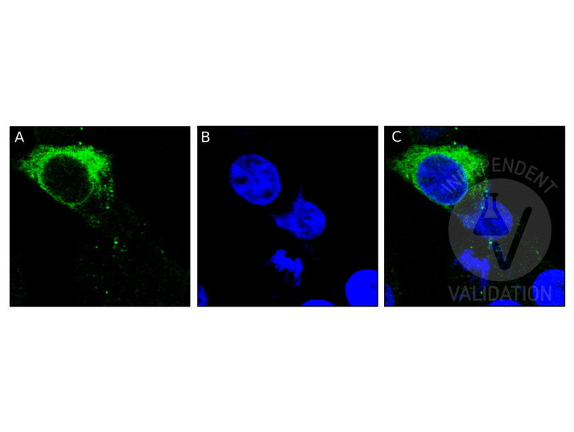
Staining with ABIN516526 shows a perinuclear pattern, suggesting MR1 localizes in the endoplasmic reticulum. No signal was detected in sample negative control tissue and the secondary antibody only control.
Validation #101752 (Immunocytochemistry)
 Validation Images
![Human MR1-expressing HEK293 cells were stained with MR1 antibody ABIN516526 and a FITC-conjugated secondary antibody (green, A). For nuclear staining, cells were stained with Hoechst (blue, B). C shows both channels merged.]() Human MR1-expressing HEK293 cells were stained with MR1 antibody ABIN516526 and a FITC-conjugated secondary antibody (green, A). For nuclear staining, cells were stained with Hoechst (blue, B). C shows both channels merged.
Protocole
Human MR1-expressing HEK293 cells were stained with MR1 antibody ABIN516526 and a FITC-conjugated secondary antibody (green, A). For nuclear staining, cells were stained with Hoechst (blue, B). C shows both channels merged.
Protocole -
- by
- Dr. Randy Brutkiewicz Laboratory, Department of Microbiology and Immunology, Indiana University School of Medicine
- No.
- #102826
- Date
- 20.02.2018
- Antigène
- MR1
- Numéro du lot
- 12045-5B5
- Application validée
- Immunoprecipitation
- Contrôle positif
- HEK293 cells transfected with human MR1 cDNA
- Contrôle négative
- HEK293 cells transfected with plasmid vector only
- Conclusion
Passed. ABIN516526 immunoprecipitates human MR1 overexpressed by HEK293 cells.
- Anticorps primaire
- ABIN516526
- Anticorps secondaire
- goat anti-rabbit Dye-IR800 conjugated antibody (Advansta, R-05060-250, lot 17083179)
- Full Protocol
- Grow HEK293 cells in DMEM medium (Lonza, 12-614F, lot 0000618582) supplemented with serum (Hyclone, SH30071.03, lot AAG205460) and antibiotics (Hyclone, SV30010, lot J150013), at 37°C and 5% CO2 dish to 70-90% confluency.
- Transfect cells with pCDNA 3.1 neo (-) (Invitrogen) containing human MR1 cDNA (Genecopoeia) using Polyethylenimine (Polysciences, 23966) following the manufacturer´s instructions.
- Lyse cells in cold lysis buffer (10mM Tris pH7.4, 150mM NaCl, 0.5mM EDTA, 2% CHAPS).
- Determine total protein content of the lysates using Commassie Protein Assay Reagent (Thermo Scientific, 1856209, lot NL179252).
- Immobilize 100µl of protein G-conjugated Sepharose beads (Pierce, product 20399, lot RI239318) ON at 4°C with
- 2.5µg mouse anti-MR1 antibody (antibodies-online, ABIN2665876, lot B177559),
- 2.5µg mouse anti-MR1 antibody (antibodies-online, ABIN516526, lot12045-5B5),
- 2.5µg rabbit anti-MR1 antibody (antibodies-online, ABIN1537116, lot SA111213CH),
- 2.5µg mouse IgG2a antibody (Biolegend, 400202, lot B153642),
- 2.5µg mouse IgG1 antibody (BD, 555746, lot 3221830), or
- 2.5µg rabbit IgG antibody (Santa Cruz Biotechnology, SC-5560, lot E0609).
- Incubate 500µg of the cell lysates with 2.5µg of antibody-bead conjugate ON at 4°C.
- Wash lysates 4x with PBS.
- Denature beads for 5min at 95°C in 60µl Laemmli SDS sample buffer and subsequently separate them on a SDS-PAGE gel using Acrylamide/Bis Premixed (Bio-Rad, 61-0125, lot 260000477) for 2-3h at 100V.
- Transfer proteins onto PVDF membrane (Millipore, IPVH00010, lot K5AA6843U) with a Western blotting system for ON at 4°C at 150mA.
- Block the membrane with blocking buffer (2% BSA/PBS/0.05%Tween-20) for 1h at RT.
- Incubate membrane with primary rabbit anti-MR1 antibody (antibodies-online ABIN1537116, lot SA111213CH) diluted 1:1000 in blocking buffer ON at 4°C.
- Wash membrane 3x for 10min with PBS/0.05%Tween-20.
- Incubate membrane with secondary goat anti-rabbit Dye-IR800 conjugated antibody (Advansta, R-05060-250, lot 17083179) diluted 1:10000 in PBS/0.05% Tween-20 for 1h at RT.
- Wash membrane 3x for 10 min with PBS/0.05% Tween-20.
- Reveal protein bands using an Odyssey imaging system (LI-COR Biosciences).
- Notes
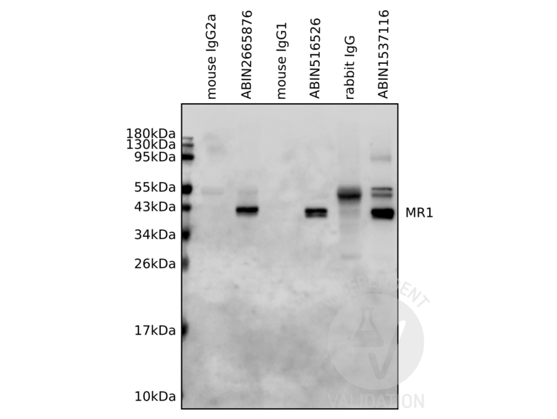
The human MR1 antibody ABIN516526, but not the isotype control, immunoprecipitates with human MR1 overexpressed by HEK293 cells.
Validation #102826 (Immunoprecipitation)
 Validation Images
![Lysates from human MR1-expressing HEK293 cells were immunoprecipitated by antibodies specific for MR1 (ABIN2665876, ABIN516526, ABIN1537116) or the respective isotype controls (mouse IgG2a, mouse, IgG1, rabbit IgG). Immunoprecipitants were resolved by SDS-PAGE gel followed by Western blotting analysis using MR1 antibody ABIN1537116.]() Lysates from human MR1-expressing HEK293 cells were immunoprecipitated by antibodies specific for MR1 (ABIN2665876, ABIN516526, ABIN1537116) or the respective isotype controls (mouse IgG2a, mouse, IgG1, rabbit IgG). Immunoprecipitants were resolved by SDS-PAGE gel followed by Western blotting analysis using MR1 antibody ABIN1537116.
Protocole
Lysates from human MR1-expressing HEK293 cells were immunoprecipitated by antibodies specific for MR1 (ABIN2665876, ABIN516526, ABIN1537116) or the respective isotype controls (mouse IgG2a, mouse, IgG1, rabbit IgG). Immunoprecipitants were resolved by SDS-PAGE gel followed by Western blotting analysis using MR1 antibody ABIN1537116.
Protocole -
- by
- Dr. Randy Brutkiewicz Laboratory, Department of Microbiology and Immunology, Indiana University School of Medicine
- No.
- #102827
- Date
- 20.02.2018
- Antigène
- MR1
- Numéro du lot
- 12045-5B5
- Application validée
- Flow Cytometry
- Contrôle positif
- HEK293 cells transfected with human MR1 cDNA
- Contrôle négative
- HEK293 cells transfected with plasmid vector only
- Conclusion
Passed. ABIN2665876 recognizes human MR1 overexpressed by HEK293 cells and can be used in flow cytometry.
- Anticorps primaire
- ABIN516526
- Anticorps secondaire
- PE-conjugated rabbit anti-mouse immunoglobulin antiserum (Jackson ImmunoResearch, 115-116-146, lot 120701)
- Full Protocol
- Grow HEK293 cells in DMEM medium (Lonza, 12-614F, lot 0000618582) supplemented with serum (Hyclone, SH30071.03, lot AAG205460) and antibiotics (Hyclone, SV30010, lot J150013), at 37°C and 5% CO2 dish to 70-90% confluency.
- Transfect cells with pCDNA 3.1 neo (-) (Invitrogen) containing human MR1 cDNA (Genecopoeia) using Polyethylenimine (Polysciences, 23966) following the manufacturer´s instructions.
- Surface staining:
- Wash 0.5x10⁶ cells 3x with HBSS/0.1% BSA.
- Incubate cells with mouse anti-MR1 antibody (antibodies-online, ABIN516526, lot 12045-5B5) diluted 1:100 or mouse anti-MR1 antibody (antibodies-online, ABIN2665876, lot B177559) diluted 1:100 for 30min on ice.
- Wash cells 3x with HBSS/0.1% BSA.
- Incubate cells with a PE-conjugated rabbit anti-mouse immunoglobulin antiserum (Jackson ImmunoResearch, 115-116-146, lot 120701) diluted 1:100 for 30min on ice.
- Total cell staining:
- Fix 0.5x10⁶ cells in 1% paraformaldehyde for 10min at RT.
- Wash cells with HBSS/BSA.
- Permeabilize cells in HBSS/BSA with 0.1% saponin for 10min at RT.
- Incubate cells with mouse anti-MR1 antibody (antibodies-online, ABIN516526, lot 12045-5B5) diluted 1:100 or mouse anti-MR1 antibody (antibodies-online, ABIN2665876, lot B177559) dilute 1:100 for 30min at RT in the presence of 0.1% saponin.
- Wash cells 3x with HBSS/0.1% BSA/0.1% saponin.
- Incubate cells with PE-conjugated rabbit anti-mouse immunoglobulin antiserum (Jackson ImmunoResearch, 115-116-146, lot 120701) diluted 1:100 for 30min at RT.
- Acquire flow cytometry data using a flow cytometry using LSR 4 (BD Biosciences) followed data analysis using FlowJo software.
- Notes
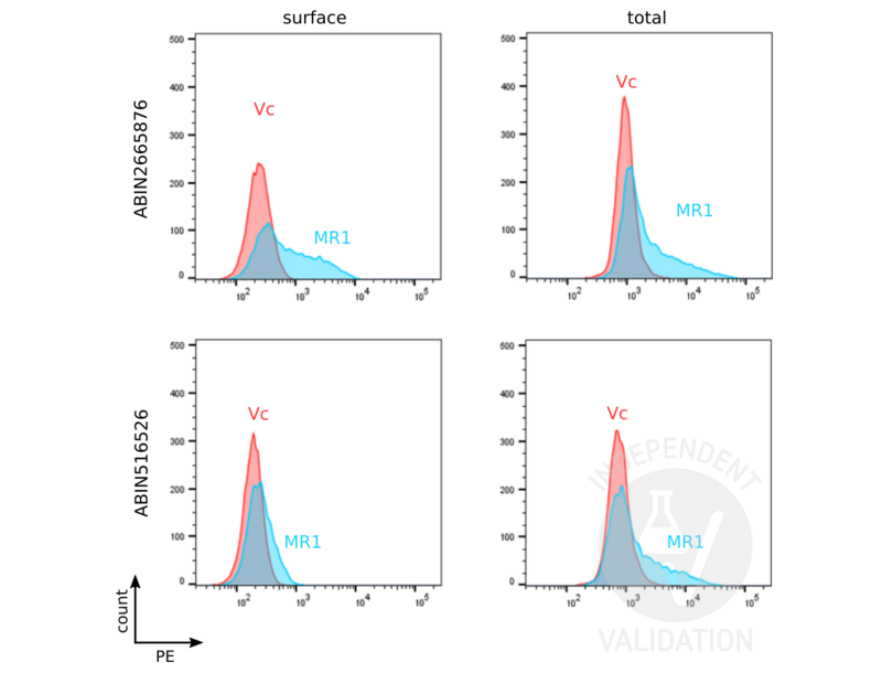
The human MR1 antibody ABIN2665876 shows specific staining for human MR1.
Validation #102827 (Flow Cytometry)
 Validation Images
![Human MR1-expressing HEK293 cells (MR1) and vector control cells (Vc) were stained with MR1 antibodies ABIN516526 or ABIN2665876 followed by PE-conjugated anti-mouse secondary antibody. For total MR1 staining, cells were permeabilized with 0.1% saponin and stained with primary and secondary antibodies. Cells were analyzed by flow cytometry.]() Human MR1-expressing HEK293 cells (MR1) and vector control cells (Vc) were stained with MR1 antibodies ABIN516526 or ABIN2665876 followed by PE-conjugated anti-mouse secondary antibody. For total MR1 staining, cells were permeabilized with 0.1% saponin and stained with primary and secondary antibodies. Cells were analyzed by flow cytometry.
Protocole
Human MR1-expressing HEK293 cells (MR1) and vector control cells (Vc) were stained with MR1 antibodies ABIN516526 or ABIN2665876 followed by PE-conjugated anti-mouse secondary antibody. For total MR1 staining, cells were permeabilized with 0.1% saponin and stained with primary and secondary antibodies. Cells were analyzed by flow cytometry.
Protocole -
-
Buffer
- In 1x PBS, pH 7.4
-
Conseil sur la manipulation
- Aliquot to avoid repeated freezing and thawing.
-
Stock
- -20 °C
-
Stockage commentaire
- Store at -20°C or lower. Aliquot to avoid repeated freezing and thawing.
-
-
- MR1 (Major Histocompatibility Complex, Class I-Related (MR1))
-
Autre désignation
- MR1
-
Sujet
-
Full Gene Name: major histocompatibility complex, class I-related
Synonyms: HLALS -
ID gène
- 3140
-
NCBI Accession
- NM_001531
-
Pathways
- Regulation of Leukocyte Mediated Immunity, Positive Regulation of Immune Effector Process, Production of Molecular Mediator of Immune Response, Cancer Immune Checkpoints
Antigène
-