CALB1 anticorps

 (2 validations)
Aperçu rapide pour CALB1 anticorps (ABIN6254097)
Antigène
Voir toutes CALB1 AnticorpsReactivité
Hôte
Clonalité
Conjugué
Application
Clone
-
-
Specificité
- Specific for endogenous levels of the ~28 kDa calbindin protein.
-
Réactivité croisée
- Boeuf (Vache), Humain, Souris, Rat
-
Attributs du produit
- Our Anti-Calbindin Antibody primary antibody is mouse monoclonal. It detects bovine, human, mouse, and rat Calbindin and is protein g purified. It is great for use in ELISA, WB, IHC.
-
Purification
- Protein G purified.
-
Immunogène
- Recombinant full length human calbindin purified from E. coli.
-
Isotype
- IgG2a
-
-
anti-Calbindin (CALB1) antibody
CALB1 Reactivité: Humain WB, IF Hôte: Lapin Monoclonal unconjugated
anti-Calbindin (CALB1) (AA 2-261) antibodyCALB1 Reactivité: Humain, Rat, Souris IHC, WB, ELISA, IF, FACS Hôte: Lapin Polyclonal unconjugated
anti-Calbindin (CALB1) (AA 2-175) antibodyCALB1 Reactivité: Humain, Rat, Souris WB, IP, IHC (p) Hôte: Lapin Polyclonal unconjugated
anti-Calbindin (CALB1) (Internal Region) antibodyVerified CALB1 Reactivité: Humain, Rat IHC, WB, ELISA Hôte: Chèvre Polyclonal unconjugated
anti-Calbindin (CALB1) (AA 7-96) antibodyCALB1 Reactivité: Humain IHC, ELISA Hôte: Souris Monoclonal CALB1-3333 unconjugated
anti-Calbindin (CALB1) (AA 2-175) antibodyCALB1 Reactivité: Humain, Rat, Souris IHC, WB, IP Hôte: Lapin Polyclonal unconjugated
anti-Calbindin (CALB1) antibodyCALB1 Reactivité: Humain, Rat, Souris, Boeuf (Vache), Poisson zèbre (Danio rerio), Poulet, Grasshopper, Singe, Non-Human Primate, Tortue IHC, WB, IP, ICC, IHC (p) Hôte: Lapin Polyclonal unconjugated
anti-Calbindin (CALB1) (AA 1-261) antibodyCALB1 Reactivité: Humain WB, ELISA Hôte: Souris Monoclonal 1F6 unconjugated
anti-Calbindin (CALB1) (AA 1-261) antibodyCALB1 Reactivité: Humain IHC, WB Hôte: Lapin Polyclonal unconjugated
anti-Calbindin (CALB1) antibodyMicroarray verified CALB1 Reactivité: Humain, Souris IHC Hôte: Souris Monoclonal MAB440307 unconjugated
-
-
Indications d'application
-
Dilution Range: WB Brain: 1:2000
Dilution Range: WB: 1:2000
-
Restrictions
- For Research Use only
-
-
- by
- Prof. Merighi, Laboratory of Neurobiology, Department of Veterinary Sciences, University of Turin
- No.
- #104494
- Date
- 02.08.2023
- Antigène
- CALB1
- Numéro du lot
- GS116G
- Application validée
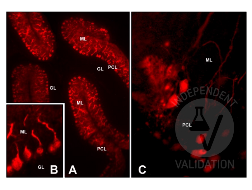
- Immunohistochemistry
- Contrôle positif
Adult (>2 months) CD1 mouse cerebellum (6 µm glass-mounted microtome sections)
Postnatal day 6-7 CD1 mouse cerebellum (cultured cerebellar slices)
- Contrôle négative
Control slices were processed for each experimental procedure, omitting the primary antibody; overnight incubation in diluent solution only.
- Conclusion
Passed. The CALB1 antibody ABIN6254097 works in IHC-P at a 1:50 dilution with or without tyramide amplification.
- Anticorps primaire
- ABIN6254097
- Anticorps secondaire
goat anti-mouse IgG (H+L) AF488-conjugated antibody (Thermo Fisher Scientific, A11034, lot 2380031)
- Full Protocol
- Indirect IMF on microtome sections
- Perfuse adult (>2 months) CD1 mice with paraformaldehyde 4% in 0.1 M phosphate buffer pH 7.4 and post-fix in the same fixative for an additional 2 h at RT.
- Wash, dehydrate, and embed samples in paraffin wax.
- Wash several times with 0.01 M PBS.
- Cut the cerebellum with a microtome into 6 µm sections and mount them on glass slides.
- After paraffin removal, incubate sections for 1 h at RT in PBS containing 1% albumin from chicken egg white (Sigma, A5378) and 0.3% Triton-X-100 (BioRad, 161-0407, lot 00583) to block non-specific binding sites.
- Incubate sections with primary mouse anti-CALB1 antibody (antibodies online, ABIN6254097, lot GS116G) diluted 1:50, 1:100, and 1:200 in 0.1 M PBS-BSA-PLL ON at RT.
- Wash 3x 5 min in 0.01 M PBS.
- Incubate sections with secondary goat anti-mouse IgG (H+L) AF488-conjugated antibody (Fisher Scientific, A11034, lot 2380031) diluted 1:500 in 0.1 M PBS for 1 h at RT.
- or alternatively with tyramide amplification:
- Incubate sections with Poly-HRP-conjugated secondary antibody for 1 h at RT.
- Wash sections 3x 5 min in 0.01 M PBS.
- Incubate sections with Tyramide working solution (for 5 sections: 5 μL 100X Tyramide stock solution, 5 μL 100X H2O2 solution, 500 µL 1X Reaction buffer) for 10 min at RT.
- Stop the reaction with the Reaction Stop Reagent working solution.
- Wash 3x 5 min in 0.01M PBS.
- Mount specimens in Fluoroshield (Sigma-Aldrich, F6182, lot MKCB0153V).
- Acquire Images with Leica DM 6000B fluorescence microscope equipped with a digital camera at 20-40x magnification.
- Indirect IMF on cultured cerebellar slices
- Euthanize CD1 mice at postnatal day 6-7 (P6-P7) with an overdose of 60 mg⁄100 g body weight sodium pentobarbital (Merck Life Science, Y0002194).
- Remove the brain removed from the skull while the head is kept submerged in ice-cooled Gey’s solution (Sigma-Aldrich) supplemented with glucose and antioxidants (for 500 mL: 4.8 mL 50% glucose, 0.05 g ascorbic acid, 0.1 g sodium pyruvate).
- Dissect the cerebellum from the brain.
- Cut 350 μm thick parasagittal slices of the cerebellum with a McIlwain tissue chopper (Brinkmann Instruments).
- Plate two to three slices onto a Millicell Cell Culture Insert (Merck Life Science, PICM0RG50).
- Place each insert inside a 35 mm Petri dish containing 1 mL of culture medium consisting of 50 % Eagle basal medium (BME, Sigma Chemicals), 25 % horse serum (Gibco by Thermo Fisher Scientific), 25 % Hanks balanced salt solution (Sigma-Aldrich), 0.5 % glucose, 0.5 % 200 mM L-glutamine, and 1ؘ % antibiotic/antimycotic solution.
- Incubate slices at 34 °C in 5 % CO2 for up to 20 d in vitro (DIV). Change the medium twice a week. Slices were allowed to equilibrate to the in vitro conditions for at least 4-6 DIV before IMF.
- Remove the culture medium from the dish and fix the slices in 1 mL of 4 % paraformaldehyde (Merck Life Science, P6148) in PBS for 1 h.
- Wash 3x 5 min in 0.01 M PBS.
- Incubate fixed cultures in PBS containing 1 % Triton X-100 (BioRad, 161-0407, lot 00583) for 10 min.
- Wash 3x 5 min in 0.01 M PBS.
- Incubate cultures ON at 4 °C under continuous stirring in PBS containing 1 % albumin from chicken egg white (Sigma, A5378) and 0.3 % Triton-X-100 (BioRad, 161-0407, lot 00583) to block non-specific binding sites.
- Incubate cultures with the primary mouse anti-CALB1 antibody (antibodies online, ABIN6254097, lot GS116G) diluted 1:50 in PBS-BSA (Sigma, A7906)-PLL (Sigma, P1524) ON at RT.
- Wash 5 x 5min in PBS.
- Incubate cultures with the secondary anti-rabbit antibody Alexa Fluor 488 diluted (Invitrogen by Thermo Fisher Scientific, A11034, lot 2380031) 1:500 in 0.1 M PBS for 1 h at RT.
- Wash 3x 5 min in 0.01 M PBS.
- Mount specimens in Fluoroshield (Sigma-Aldrich, F6182, lot MKCB0153V).
- Acquire Images with Leica DM 6000B fluorescence microscope equipped with a digital camera at 20-40x magnification.
- Notes
For indirect IMF on cerebellum paraffin sections, antigen retrieval treatment was also tested. In this case, sections were processed for microwave antigen retrieval for 10 min (95-100 °C) in 10 mM sodium citrate buffer (pH 6.0). After 20 min of spontaneous cooling, sections were washed twice for 5 min with distilled water and twice for 5 min with PBS.
Validation #104494 (Immunohistochemistry)
 Validation Images
![]() Protocole
Protocole -
- by
- Prof. Merighi, Laboratory of Neurobiology, Department of Veterinary Sciences, University of Turin
- No.
- #104530
- Date
- 02.08.2023
- Antigène
- CALB1
- Numéro du lot
- GS116G
- Application validée
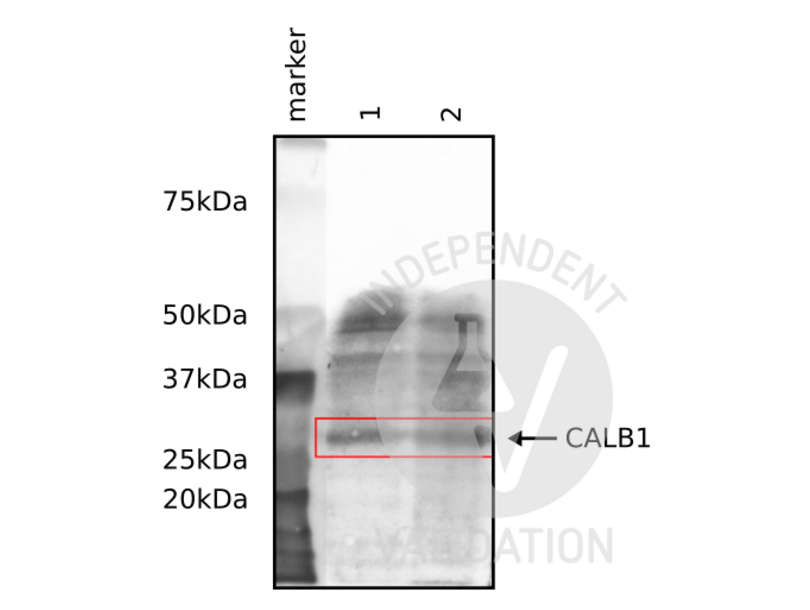
- Western Blotting
- Contrôle positif
Adult mouse brain and cerebellum
- Contrôle négative
- Conclusion
Passed. The CALB1 antibody ABIN6254097 works in WB at a 1:1000 dilution.
- Anticorps primaire
- ABIN6254097
- Anticorps secondaire
goat anti-mouse IgG (H+L) HRP-conjugated (Thermo Fisher Scientific, G-21040)
- Full Protocol
- Homogenize tissues with cold lysis buffer containing 50 mM Tris HCl, 150 mM NaCl, 1% Triton X-100, 1 mM EDTA, and 1% protease inhibitor (Sigma P8340) using an ultrasonic homogenizer (MSE, SoniPrep 150) with 16 amplitude, 20 s on, 10 s off pulse for 60 s.
- Centrifuge tissue homogenates at 13,000 rpm for 20 min at 4 °C.
- Collect supernatants and determine total protein content using a Bradford assay.
- Denature 100 µg of total protein for 5 min at 90 °C and subsequently separate them on a denaturing 12% PAGE-SDS gel alongside a Precision Plus Protein Dual Color Standard (Bio-Rad, 160374).
- Electro-transfer proteins onto nitrocellulose membrane (Amersham Biosciences, RPN203D) ON in the cold room.
- Wash membrane 3x for 10 min with 0.01 M PBS containing 0.1% Tween-20 (PBST).
- Block membrane with PBST containing 2% bovine serum albumin for 1 h at RT.
- Incubate membrane with primary rabbit anti-CALB1 antibody (antibodies-online, ABIN6254097, lot GS116G) diluted 1:1,000 in PBST ON at 4 °C.
- Wash membrane 3x 10 min with PBST.
- Incubate membrane with secondary HRP-conjugated goat anti-mouse IgG (Thermo Fisher Scientific, G-21040) diluted 1:50,000 in PBST for 1 h at RT.
- Wash membrane 3x 10 min with PBST.
- Visualize proteins with SuperSignal West Atto Ultimate Sensitivity Substrate (Thermo Fisher Scientific, A38555) using a ChemiDoc Imaging System.
- Notes
Validation #104530 (Western Blotting)
 Validation Images
![Western blot results using ABIN6254097 to reveal CALB1 (28 kDa) in the adult mouse brain and cerebellum.]() Western blot results using ABIN6254097 to reveal CALB1 (28 kDa) in the adult mouse brain and cerebellum.
Protocole
Western blot results using ABIN6254097 to reveal CALB1 (28 kDa) in the adult mouse brain and cerebellum.
Protocole -
-
Format
- Liquid
-
Buffer
- PBS + 50 % glycerol and 5 mM Sodium azide
-
Agent conservateur
- Sodium azide
-
Précaution d'utilisation
- This product contains Sodium azide: a POISONOUS AND HAZARDOUS SUBSTANCE which should be handled by trained staff only.
-
Stock
- -20 °C
-
Stockage commentaire
- Storage at -20°C is recommended, as aliquots may be taken without freeze/thawing due to presence of 50% glycerol. Stable for at least 1 year at -20°C.
-
Date de péremption
- 12 months
-
-
- CALB1 (Calbindin (CALB1))
-
Autre désignation
- CALB1
-
Sujet
-
Synonyms: avian-type antibody, CAB27 antibody, CALB 1 antibody, CALB antibody, CALB1 antibody, CALB1_HUMAN antibody, Calbindin 1 28 kDa antibody, Calbindin antibody, Calbindin D28 antibody, D 28K antibody, D-28K antibody, D28K antibody, OTTHUMP00000166027 antibody, OTTHUMP00000225441 antibody, RTVL H protein antibody, Vitamin D dependent calcium binding protein antibody, Vitamin D dependent calcium binding protein avian type antibody, Vitamin D-dependent calcium-binding protein antibody
Description Logic: Our Anti-Calbindin Antibody primary antibody is mouse monoclonal. It detects bovine, human, mouse, and rat Calbindin and is protein g purified. It is great for use in ELISA, WB, IHC.
Gene Name Alternatives: CALB1
-
Poids moléculaire
- 28 kDa
-
ID gène
- 793
-
UniProt
- P05937
Antigène
-