RFP anticorps
 (1 reference)
 (1 validation)
Aperçu rapide pour RFP anticorps (ABIN967350)
Antigène
Voir toutes RFP AnticorpsReactivité
Hôte
Clonalité
Conjugué
Application
Clone
-
-
Attributs du produit
-
1. Since applications vary, each investigator should titrate the reagent to obtain optimal results.
2. Please refer to us for technical protocols.
3. Caution: Sodium azide yields highly toxic hydrazoic acid under acidic conditions. Dilute azide compounds in running water before discarding to avoid accumulation of potentially explosive deposits in plumbing.
4. Source of all serum proteins is from USDA inspected abattoirs located in the United States. -
Purification
- The monoclonal antibody was purified from tissue culture supernatant or ascites by affinity chromatography.
-
Immunogène
- DsRed1 recombinant protein
-
Isotype
- IgG1
-
-
anti-Red Fluorescent Protein (RFP) antibody
RFP Reactivité: Discosoma WB, ELISA, IF, IHC, IP, FACS, IHC (fro), IHC (p) Hôte: Lapin Polyclonal unconjugated
anti-Red Fluorescent Protein (RFP) antibodyRFP Reactivité: Discosoma WB, ELISA, IP, FACS, FM Hôte: Souris Monoclonal 8E5-G7 unconjugated
anti-Red Fluorescent Protein (RFP) antibodyRFP Reactivité: Discosoma, Tomato WB, IF, IHC (fro), IHC (p), IEM Hôte: Chèvre Polyclonal unconjugated
anti-Red Fluorescent Protein (RFP) antibodyRFP Reactivité: Discosoma WB, ELISA Hôte: Poulet Polyclonal unconjugated
anti-Red Fluorescent Protein (RFP) antibody (DyLight 550)RFP Reactivité: Discosoma, Tomato WB, IF, IHC (fro) Hôte: Chèvre Polyclonal DyLight 550
anti-Red Fluorescent Protein (RFP) antibody (DyLight 488)RFP Reactivité: Discosoma, Tomato WB, IF, IHC (fro) Hôte: Chèvre Polyclonal DyLight 488
anti-Red Fluorescent Protein (RFP) antibody (DyLight 633)RFP Reactivité: Discosoma, Tomato WB, IF, IHC (fro) Hôte: Chèvre Polyclonal DyLight 633
anti-Red Fluorescent Protein (RFP) antibody (Biotin)RFP Reactivité: Discosoma WB, ELISA, IHC Hôte: Lapin Polyclonal Biotin
anti-Red Fluorescent Protein (RFP) antibody (Biotin)RFP Reactivité: Discosoma WB, ELISA, IF, FM Hôte: Poulet Polyclonal Biotin
anti-Red Fluorescent Protein (RFP) antibody (DyLight 405)RFP Reactivité: Discosoma, Tomato WB, IF, IHC (fro) Hôte: Chèvre Polyclonal DyLight 405
-
-
Indications d'application
- Applications include western blot analysis (0.25-1.0 µg/ml). DsRed2 control lysate is provided as a positive control (component 51-16616N) and approximately 10 µl is recommended to visualize DsRed by western blot analysis.
-
Commentaires
-
Related Products: ABIN967389
-
Restrictions
- For Research Use only
-
-
- by
- Institut für Virologie, Medizinische Hochschule Hannover
- No.
- #101231
- Date
- 19.06.2017
- Antigène
- DsRed
- Numéro du lot
- 6223801
- Application validée
- Western Blotting
- Contrôle positif
- Human foreskin fibroblasts infected with DsRed expressing virus
- Contrôle négative
- Cells infected with EGFP expressing virus
- Conclusion
- Passed. ABIN967350 specifically recognizes DsRed in extracts of cells expressing DsRed and GFP.
- Anticorps primaire
- ABIN967350
- Anticorps secondaire
- goat anti-mouse antibody (Pierce, 32230, lot QC212446)
- Full Protocol
- Grow fibroblasts in Dulbecco’s modified Eagle’s medium (Biochrom, F-0435, lot 0315F) supplemented with 10% fetal calf serum (FBS Good Forte, PAN Biotech, P40-47500, lot P130912)and 100U/ml penicillin, 100µg/ml streptomycin sulfate (PAN Biotech, P06-19100, lot 7530716) at 37°C and 5% CO2 in 2ml on a tissue culture 6-well plate to 90% confluency.
- Infect cells at an MOI of 3 with
- an EGFP-expressing virus V1-gfp,
- a DsRed expressing viruse V2-DsRed,
- a DsRed expressing viruse V3-DsRed,
- virus V1-gfp and V2-DsRed, or
- virus V1-gfp and V3-DsRed
- Grow cells for 4d at 37°C and 5% CO2.
- Collect 8.5x10⁴ cells directly in 200µl of1x Laemmli SDS sample buffer, and boil them for 5min at 95°C.
- Separate the equivalent of 5000 cells of each sample on an Any kD SDS-PAGE gel (Bio-Rad, 456-9036, control 400100025) in a Bio-Rad Mini-Protean gel electrophoresis chamber for 45min at 250V and 21°C.
- Transfer proteins onto nitrocellulose membrane (GE Healthcare, 10600003, lot G7724136) with a TE77X semi-dry blotting system (SERVA) for 30min at 0.5A.
- Block the membrane using Roti-Block solution (Carl Roth, A151.1, lot 382190165) for 1h at RT.
- Incubate the membrane with primary
- mouse anti-Red Fluorescent Protein (DsRed) antibody (antibodies-online, ABIN967350, lot 6223801)
- rabbit anti-GFP (D5.1) XP (Cell Signalling, 2956, lot 4) antibody
- diluted 1:1000 in Roti-Block solution ON at RT.
- Wash the membrane 4x for 5min in PBS + 0.1% Tween20 (PBST).
- Incubate the membrane with secondary HRP-conjugated
- goat anti-mouseantibody (Pierce, 32230, lot QC212446)
- goat anti-rabbitantibody (Pierce, 32260, lot QH219992)
- diluted 1:2000 in PBST for 1h at RT.
- Wash the membrane 4x for 5min in PBST.
- Visualize protein bands using SuperSignal West Femto Maximum Sensitivity Substrate (Thermo Scientific, 34096, lot RH236205) on a LAS 3000 imager (Fuji).
- Strip the membrane using Restore PLUS Western blot stripping buffer (Thermo Scientific, 46428, lot PA196499) for 20min at RT.
- Wash the membrane 4x for 5min in PBST, and block for 1h at RT with Roti-Block solution.
- Incubate the membrane with primary rabbit anti-GAPDH antibody (Cell signalling, 2118, lot 10) diluted 1:2000 in PBST for 1h at RT.
- Wash the membrane 4x for 5min in PBST.
- Incubate the membrane with secondary goat anti-rabbit antibody (PIERCE, 32260, lot QH219992) diluted 1:2000 in PBST for 1h at RT.
- Wash the membrane 4x for 5min in PBST.
- Visualize protein bands using SuperSignal West Femto Maximum Sensitivity Substrate (Thermo Scientific, 34096, lot RH236205) on a LAS 3000 imager (Fuji).
- Notes
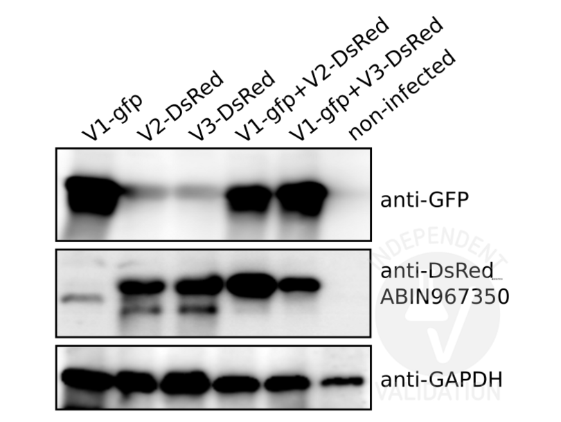
The Red Fluorescent Protein (DsRed) antibody ABIN967350 reveals a protein of the expected molecular weight (31kDa) in lysates of cells infected with DsRed or with DsRed and EGFP expressing virus. A slightly lower MW band is visible in the lysates of EGFP (expected MW 28kDa) expressing cells. This might reflect some cross-reactivity of the DsRed antibody with the structurally similar EGFP. However, the overall selectivity of ABIN967350 towards DsRed is very good.
Validation #101231 (Western Blotting)
 Validation Images
![Immunoblot of DsRed or EGFP expressing human fibroblast lysates using ABIN967350. Cells were infected with EGFP-expressing virus (V1-gfp), DsRed-expressing viruses (V2-/V3-DsRed), or co-infected as indicated. The lane on the far right contains a sample from non-infected cells.]() Immunoblot of DsRed or EGFP expressing human fibroblast lysates using ABIN967350. Cells were infected with EGFP-expressing virus (V1-gfp), DsRed-expressing viruses (V2-/V3-DsRed), or co-infected as indicated. The lane on the far right contains a sample from non-infected cells.
Protocole
Immunoblot of DsRed or EGFP expressing human fibroblast lysates using ABIN967350. Cells were infected with EGFP-expressing virus (V1-gfp), DsRed-expressing viruses (V2-/V3-DsRed), or co-infected as indicated. The lane on the far right contains a sample from non-infected cells.
Protocole -
-
Format
- Liquid
-
Concentration
- 0.25 mg/mL
-
Buffer
- Aqueous buffered solution containing BSA, glycerol, and ≤0.09 % sodium azide.
-
Agent conservateur
- Sodium azide
-
Précaution d'utilisation
- This product contains Sodium azide: a POISONOUS AND HAZARDOUS SUBSTANCE which should be handled by trained staff only.
-
Stock
- -20 °C
-
Stockage commentaire
- Store undiluted at -20°C.
-
-
-
: "The structural basis for red fluorescence in the tetrameric GFP homolog DsRed." dans: Nature structural biology, Vol. 7, Issue 12, pp. 1133-8, (2000) (PubMed).
-
: "The structural basis for red fluorescence in the tetrameric GFP homolog DsRed." dans: Nature structural biology, Vol. 7, Issue 12, pp. 1133-8, (2000) (PubMed).
-
- RFP (Red Fluorescent Protein (RFP))
-
Autre désignation
- DsRed Fluorescent Protein
-
Classe de substances
- Chemical
-
Sujet
- DsRed is naturally occurring fluorescent protein from a coral of the genus Discosoma. DsRed, like green fluorescent protein (GFP) can be introduced into either cultured cells or in transgenic animals to study the localization of a specific protein. It can also be used in fusion constructs with a protein of interest to examine protein-protein interactions or as a general marker. The crystal structure of DsRed has been solved and shown that DsRed monomers exhibit similar topology to GFP, but have additional chemical modifications which account for differences in the spectra, when compared to GFP. DsRed has a longer emission/absorption wavelength than GFP. DsRed migrates at 31 kDa in SDS/PAGE. The antibody recognizes three forms of DsRed, 1, 2 and pTimer. A recombinant protein representing DsRed1 was used as the immunogen.
-
Poids moléculaire
- 31 kDa
Antigène
-


