TDC2 anticorps (C-Term)
 (1 validation)
Aperçu rapide pour TDC2 anticorps (C-Term) (ABIN4889606)
Antigène
Reactivité
Hôte
Clonalité
Application
-
-
Épitope
- C-Term
-
Specificité
- Reacts with Drosophila melanogaster 72 kDa Tdc2 protein
-
Purification
- Purified (protein A)
-
Immunogène
- Synthetic peptide derived from C-terminal part of Drosophila Tdc2 protein.
-
-
-
Indications d'application
-
Working dilution: Optimal dilutions should be determined by the end user.
The following are guidelines only:
IHC(1:200 - 1:1000) WB(1:200 - 1:2000) -
Restrictions
- For Research Use only
-
-
- by
- Department of Entomology, University of California, Riverside
- No.
- #100812
- Date
- 26.05.2017
- Antigène
- Tdc2
- Numéro du lot
- 13B1
- Application validée
- Immunofluorescence
- Contrôle positif
D. melanogaster octopaminergic neurons labeled with Tdc2-Gal4 of the abdominal nerve to the ovary
- Contrôle négative
- Conclusion
Passed. ABIN809182 labels octopaminergic neurons specifically and with no background.
- Anticorps primaire
- ABIN4889606
- Anticorps secondaire
- goat anti-rabbit AF542 conjugated antibody (Life Technologies)
- Full Protocol
- Dissect ovaries of D. melanogaster ETHR-Gal4/UAS-MCD8-GFP expressing GFP in octopaminergic neurons in cold Schneider’s Insect Medium (S2; Sigma Aldrich, S01416).
- Transfer tissue to 2ml protein LoBind tubes (Eppendorf, 022431102) filled with S2 containing 2% paraformaldehyde (PFA) at RT.
- Fix tissue for 55min at RT while nutating.
- Wash tissue 4x 10min with 1.75ml PBS containing 0.5% Triton X-100 (PBST).
- Remove PBST and add 200µl 5% goat serum (GS; Thermo Fisher Scientific, 16210064) in PBST per tube.
- Incubate 1.5h at RT on a rotator.
- Remove blocking solution.
- Incubate with primary
- rabbit anti-Tdc2 antibody (Tyrosine Decarboxylase 2) (C-Term) (antibodies-online, ABIN4889606, lot 13B1) diluted 1:200 in blocking solution.
- mouse anti-GFP (Thermo Fisher Scientific) diluted 1:500 in blocking solution.
- Incubate for 4h at RT followed by 36-48h at 4°C on a rotator.
- Rinse tissue with 1.75ml PBST. Allow the tissue to settle to the bottom before removing the liquid.
- Wash tissue 3x 30min with 1.75ml PBST.
- Incubate with secondary 200µl secondary goat anti-rabbit AF542 conjugated antibody (Life Technologies) and goat anti-mouse AF488 conjugated antibody (Life Technologies, A11034) diluted 1:500 in blocking solution containing 0.5mg/ml DAPI.
- Incubate for 4h at RT followed by 72h at 4°C on a rotator.
- Rinse tissue with 1.75ml PBST. Allow the tissue to settle to the bottom before removing the liquid.
- Wash tissue 3x 30min with 1.75ml PBST.
- Add 1.75ml PBST containing 4% PFA at RT.
- Fix tissue for 5h at RT while nutating.
- Rinse tissue with 1.75ml PBST. Allow the tissue to settle to the bottom before removing the liquid.
- Wash tissue 4x 15min with 1.75ml PBST.
- Mount tissue on a poly-L-lysine (Sigma Aldrich, P1524-25MG) coated cover glass.
- Dehydrate tissue by covering the cover glass for 10min each with 30%, 50%, 75%, 95%, 100%, 100%, and 100% EtOH.
- Clearing using by covering the cover glass 3x 5min with xylene.
- Add 7 drops of dibutyl phthalate in xylene (DPX) on top of the mounted tissue.
- Seat cover glass face down gently onto a prepared slide with spacers.
- Let the slide dry for 48h at RT in a hood before viewing.
- Notes
Staining of ETHR-Gal4/UAS-MCD8-GFP expressing GFP in octopaminergic neurons was performed as previously described.
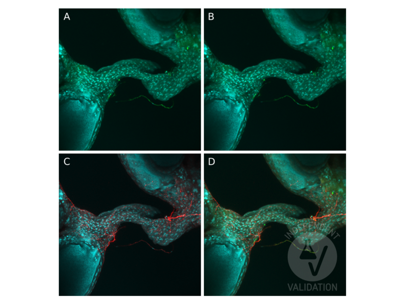
ABIN4889606 worked fantastically. It labeled octopaminergic neurons specifically and with no background. Pictures are below. The well-characterized neurons labeled with Tdc2-Gal4 of the abdominal nerve to the ovary described in Middleton et al. (2006) overlapped with ABIN4889606. Staining with ABIN4889606 was stronger than Gal4 labeling.
Validation #100812 (Immunofluorescence)
 Validation Images
![Immunostaining of D. melanogaster octopaminergic ovarian nerves expressing GFP (A), immunostaining of expressed GFP (green) (B), and of tdc2 with ABIN4889606 (red) (C). The three channels are merged in D.]() Immunostaining of D. melanogaster octopaminergic ovarian nerves expressing GFP (A), immunostaining of expressed GFP (green) (B), and of tdc2 with ABIN4889606 (red) (C). The three channels are merged in D.
Protocole
Immunostaining of D. melanogaster octopaminergic ovarian nerves expressing GFP (A), immunostaining of expressed GFP (green) (B), and of tdc2 with ABIN4889606 (red) (C). The three channels are merged in D.
Protocole -
-
Format
- Lyophilized
-
Reconstitution
- Must be reconstituted in distilled water.
-
Concentration
- 1 mg/mL
-
Buffer
- Tris 0,1M, glycine 0,1M, sucrose 2 %
-
Stock
- 4 °C/-20 °C
-
Stockage commentaire
- Lyophilized powder stable for a minimum of 2 years at -20°C. Store reconstituted antibodies at +4°C. For extended periods store in aliquots at -20°C. Antibodies are guaranteed for 6 month from date of receipt.
-
Date de péremption
- 24 months
-
-
- TDC2 (Tyrosine Decarboxylase 2 (TDC2))
-
Autre désignation
- Tyrosine Decarboxylase 2
-
Sujet
- Enzyme involved in tyrosine metabolism. Use Pyridoxal phosphate as cofactor.
-
ID gène
- 246620
-
UniProt
- A1Z6N4
Antigène
-


