TUBB3 anticorps (C-Term)
 (1 reference)
 (1 validation)
Aperçu rapide pour TUBB3 anticorps (C-Term) (ABIN1742553)
Antigène
Voir toutes TUBB3 AnticorpsReactivité
Hôte
Clonalité
Conjugué
Application
-
-
Épitope
- C-Term
-
Fonction
- Rabbit anti-beta3-Tubulin antiserum
-
Specificité
- Specific for beta3-tubulin.
-
Réactivité croisée (Details)
- Reacts with: human (Q13509), rat (Q4QRB4), mouse (Q9ERD7). Other species not tested yet.
-
Purification
- antiserum
-
Immunogène
- Synthetic peptide corresponding to residues near the carboxy terminus of mouse beta3-tubulin (UniProt Id: Q9ERD7)
-
-
anti-Tubulin, beta 3 (TUBB3) (N-Term) antibody
TUBB3 Reactivité: Humain, Souris, Rat, Chien, Porc WB, ICC, IHC (p), ICFC Hôte: Souris Monoclonal TU-20 unconjugated
anti-Tubulin, beta 3 (TUBB3) (N-Term) antibody (FITC)TUBB3 Reactivité: Humain, Souris, Rat, Chien, Porc FACS, ICC Hôte: Souris Monoclonal TU-20 FITC
anti-Tubulin, beta 3 (TUBB3) (AA 36-63) antibodyTUBB3 Reactivité: Humain, Souris WB, FACS, IHC (p) Hôte: Lapin Polyclonal RB21098 unconjugated
anti-Tubulin, beta 3 (TUBB3) antibodyTUBB3 Reactivité: Humain WB, IF, IP Hôte: Lapin Monoclonal unconjugated
anti-Tubulin, beta 3 (TUBB3) (AA 36-63) antibodyTUBB3 Reactivité: Humain, Souris WB, IHC, ELISA, FACS Hôte: Lapin Polyclonal unconjugated
anti-Tubulin, beta 3 (TUBB3) antibodyTUBB3 Reactivité: Humain, Souris, Rat ELISA, FACS, ICC Hôte: Souris Monoclonal 2E9 unconjugated
anti-Tubulin, beta 3 (TUBB3) antibodyTUBB3 Reactivité: Humain WB, IHC, ELISA Hôte: Souris Monoclonal unconjugated
anti-Tubulin, beta 3 (TUBB3) antibodyTUBB3 Reactivité: Humain WB, IHC, ELISA, IF, IP Hôte: Souris Monoclonal unconjugated
anti-Tubulin, beta 3 (TUBB3) (AA 1-210) antibodyTUBB3 Reactivité: Humain WB, IHC, ELISA Hôte: Lapin Polyclonal unconjugated
anti-Tubulin, beta 3 (TUBB3) (AA 1-210) antibodyTUBB3 Reactivité: Humain WB, IHC, ELISA, IF Hôte: Lapin Polyclonal unconjugated
-
-
Indications d'application
- WB:1 : 1000 up to 1 : 10000 , IP:yes ICC:1 : 1000 up to 1 : 5000, IHC:1 : 200 up to 1 : 500, IHC-P:1 : 500,
-
Restrictions
- For Research Use only
-
-
- by
- Kinexus Bioinformatics Corporation
- No.
- #029822
- Date
- 28.10.2014
- Antigène
- Numéro du lot
- 302302/3
- Application validée
- Western Blotting
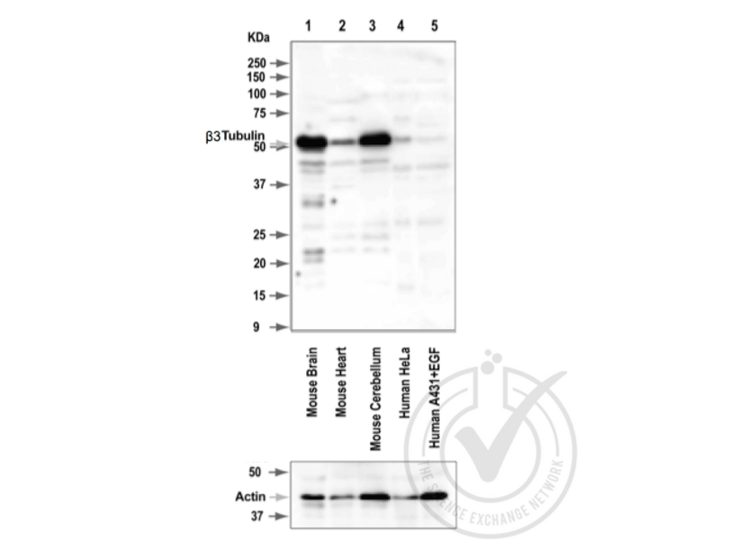
- Contrôle positif
- Mouse brain
- Contrôle négative
- Mouse heart, HeLa cells, A431 cells
- Conclusion
- A band was observed in the positive controls at the expected size (~50 kDa), which is lower in the negative controls.
- Anticorps primaire
- Antigen: Tubulin, Beta, 3 (TUBB3) (AA 443-450)
- Catalog number: ABIN1742553
- Lot number: 302302/3
- Dilution: 1:1000
- Anticorps secondaire
- Antibody: Donkey anti-Rabbit IgG Antibody (HRP)
- Lot number: F0613
- Dilution: 1:10,000
- Full Protocol
- 1. Cell/tissue total protein lysates were boiled in 1X SDS Sample Buffer containing 1% SDS and 1.25% β-mercaptoethanol at 95°C for 5 minutes prior to loading.
- 2. 15 μg of boiled lysate were loaded and resolved on a 12% SDS-polyacrylamide gel.
- 3. The Precision Plus Protein™ All Blue Prestained Standards from BioRad (161-0373) were used as molecular mass markers.
- 4. Proteins were transferred onto nitrocellulose membrane by tank transfer and protein transfer was confirmed with Ponceau S staining.
- 5. The immunoblot membrane was blocked in 2.5% skim milk and 1.5% BSA solution in TTBS at room temperature for 60 minutes.
- 6. The membrane was washed in TTBS twice for 5 minutes each.
- 7. The membrane was immersed with the protein side up in the antibody solution in TBS and incubated overnight at 4°C with gentle agitation.
- 8. The membrane was rinsed twice with TTBS.
- 9. The membrane was washed in TTBS twice for 5 minutes each.
- 10. The membrane was washed in TTBS once for 15 minutes.
- 11. The membrane was incubated in the HRP-conjugated secondary antibody solution in TBS for 60 minutes at room temperature with gentle agitation.
- 12. The membrane was rinsed twice with TTBS.
- 13. The membrane was washed in TTBS twice for 5 minutes each.
- 14. The membrane was washed in TTBS once for 15 minutes.
- 15. Signals were detected by chemiluminescence (ECL). The blot was scanned for 320 seconds.
- 16. The membrane was rinsed three times with TTBS.
- 17. Repeated Steps 4-15 with the loading control antibody and its matching secondary antibody. The blot was scanned for 160 seconds.
- Notes
- - No experimental challenges noted.
Validation #029822 (Western Blotting)
 Validation Images
![Figure 1: Western Blot for Tubulin, Beta, 3 (TUBB3). Grey arrowhead indicates the expected molecular weight of ~50 kDa.]() Figure 1: Western Blot for Tubulin, Beta, 3 (TUBB3). Grey arrowhead indicates the expected molecular weight of ~50 kDa.
Protocole
Figure 1: Western Blot for Tubulin, Beta, 3 (TUBB3). Grey arrowhead indicates the expected molecular weight of ~50 kDa.
Protocole -
-
Format
- Lyophilized
-
Reconstitution
- For reconstitution add 200 μLH2O, then aliquot and store at -20 °C until use.
-
Conseil sur la manipulation
- Do not freeze! Antibodies should be stored at +4°C when still lyophilized.
-
Stock
- -20 °C,4 °C
-
Stockage commentaire
- Antibodies should be stored at +4°C when still lyophilized. Do not freeze!
-
-
-
: "Neuron-astrocyte interaction enhance GABAergic synaptic transmission in a manner dependent on key metabolic enzymes." dans: Frontiers in cellular neuroscience, Vol. 9, pp. 120, (2015) (PubMed).
-
: "Neuron-astrocyte interaction enhance GABAergic synaptic transmission in a manner dependent on key metabolic enzymes." dans: Frontiers in cellular neuroscience, Vol. 9, pp. 120, (2015) (PubMed).
-
- TUBB3 (Tubulin, beta 3 (TUBB3))
-
Autre désignation
- beta3-Tubulin
-
Sujet
-
Synonyms: TuJ1, TUBB3, Class III ß-Tubulin, ßIII-Tubulin, betaIII-Tubulin, eye-retina
Antigen Information: beta3-Tubulin also known as TuJ1 is a neuron specific Tubulin
-
UniProt
- Q9ERD7
-
Pathways
- Dynamique des Microtubules, M Phase
Antigène
-


