RAD21 anticorps (Center)
 (2 références)
 (1 validation)
Aperçu rapide pour RAD21 anticorps (Center) (ABIN2856242)
Antigène
Voir toutes RAD21 AnticorpsReactivité
Hôte
Clonalité
Conjugué
Application
-
-
Épitope
- Center
-
Attributs du produit
-
Rabbit Polyclonal antibody to RAD21 (RAD21 homolog (S. pombe))
RAD21 antibody -
Purification
- Purified by antigen-affinity chromatography.
-
Immunogène
- Recombinant protein encompassing a sequence within the center region of human RAD21. The exact sequence is proprietary.
-
Isotype
- IgG
-
-
anti-RAD21 Homolog (RAD21) (N-Term) antibody
RAD21 Reactivité: Humain, Souris WB, IF Hôte: Souris Monoclonal unconjugated Recombinant Antibody
anti-RAD21 Homolog (RAD21) antibodyRAD21 Reactivité: Humain WB, IHC, IF, IP, ChIP Hôte: Lapin Polyclonal unconjugated
anti-RAD21 Homolog (RAD21) antibodyKD Validated RAD21 Reactivité: Humain WB, FACS, ICC Hôte: Lapin Monoclonal 24GB1390 unconjugated Recombinant Antibody
anti-RAD21 Homolog (RAD21) (AA 287-403) antibodyRAD21 Reactivité: Humain, Rat, Singe WB, ELISA, IHC, FACS, ICC Hôte: Souris Monoclonal 1B6D1 unconjugated
anti-RAD21 Homolog (RAD21) (C-Term) antibodyRAD21 Reactivité: Humain, Souris WB, ELISA, IHC, IF, ICC Hôte: Lapin Polyclonal unconjugated
anti-RAD21 Homolog (RAD21) antibodyRAD21 Reactivité: Humain WB, ChIP Hôte: Lapin Monoclonal unconjugated
anti-RAD21 Homolog (RAD21) (AA 373-422) antibodyRAD21 Reactivité: Humain, Souris, Rat, Boeuf (Vache), Lapin, Chien, Cheval, Singe, Cobaye, Poulet, Roussette (Chauve-souris), Porc, Xenopus laevis WB Hôte: Lapin Polyclonal unconjugated
anti-RAD21 Homolog (RAD21) (C-Term) antibodyRAD21 Reactivité: Humain, Souris, Rat, Boeuf (Vache), Poisson zèbre (Danio rerio) WB, IHC, IF, IC Hôte: Lapin Polyclonal unconjugated
anti-RAD21 Homolog (RAD21) (C-Term) antibodyRAD21 Reactivité: Humain, Souris, Hamster WB, IF Hôte: Lapin Polyclonal unconjugated
anti-RAD21 Homolog (RAD21) (AA 531-631) antibodyRAD21 Reactivité: Humain, Rat WB, ELISA, FACS, IF (cc), IF (p), IHC (p), IHC (fro) Hôte: Lapin Polyclonal unconjugated
-
-
Indications d'application
- Suggested dilution Reference ICC/IF 1:100-1:1000* IHC (Formalin-fixed paraffin-embedded sections) 1:100-1:1000* Immunoprecipitation 1:100-1:500* Western blot 1:500-1:3000* Not tested in other applications. *Optimal dilutions/concentrations should be determined by the researcher.Suggested dilutionReferenceICC/IF1:100-1:1000* IHC (Formalin-fixed paraffin-embedded sections)1:100-1:1000* Immunoprecipitation1:100-1:500* Western blot1:500-1:3000*
-
Commentaires
-
Positive Control: K562 , THP-1 , HL-60 , NIH-3T3 , JC , BCL-1
-
Restrictions
- For Research Use only
-
-
- by
- Cantù Lab, Gene Regulation during Development and Disease, Linköping University
- No.
- #104616
- Date
- 06.12.2024
- Antigène
- RAD21
- Numéro du lot
- Application validée
- Cleavage Under Targets and Release Using Nuclease
- Contrôle positif
- Anti H3K4me (antibodies-online, ABIN3023251)
- Contrôle négative
- Guinea Pig anti-rabbit IgG (antibodies-online, ABIN101961)
- Conclusion
Passed.
- Anticorps primaire
- ABIN2856242
- Anticorps secondaire
- Full Protocol
- Cell harvest and nuclear extraction
- Harvest 250,000 HEK293T cells per antibody
- Centrifuge cell solution 5 min at 600 x g at RT.
- Remove the liquid carefully.
- Gently resuspend cells in 2 mL of Nuclear Extraction Buffer (20 mM HEPES-KOH pH 8.2, 20% Glycerol, 0,05% IGEPAL, 0.5 mM Spermidine, 10 mM KCl, Roche Complete Protease Inhibitor EDTA-free).
- Move the solution to a 2 mL centrifuge tube.
- Pellet the nuclei 800 x g for 5 min.
- Repeat the NE wash twice for a total of three washes.
- Resuspend the nuclei in 40 µL NE Buffer per sample.
- Concanavalin A beads preparation
- Prepare one 2 mL microcentrifuge tube.
- Gently resuspend the magnetic Concanavalin A Beads (antibodies-online, ABIN6923139).
- Pipette 10 µL Con A Beads slurry for each sample into the 2 mL microcentrifuge tube.
- Place the tube on a magnet stand until the fluid is clear. Remove the liquid carefully.
- Remove the microcentrifuge tube from the magnetic stand.
- Pipette 1 mL Binding Buffer (20 mM HEPES pH 7.5, 10 mM KCl, 1 mM CaCl2, 1 mM MnCl2) into the tube and resuspend ConA beads by gentle pipetting.
- Spin down the liquid from the lid with a quick pulse in a table-top centrifuge.
- Place the tubes on a magnet stand until the fluid is clear. Remove the liquid carefully.
- Remove the microcentrifuge tube from the magnetic stand.
- Repeat the wash twice for a total of three washes.
- Gently resuspend the ConA Beads in 44ul binding buffer
- Nuclei immobilization – binding to Concanavalin A beads
- Carefully vortex the nuclei suspension and add 44 µL of the Con A beads in Binding Buffer to the cell suspension for each sample.
- Close tube tightly incubates 10 min at 4 °C.
- Put the 2 mL tube on the magnet stand and when the liquid is clear remove the supernatant.
- Resuspend the beads in 1 mL of EDTA wash buffer (20 mM HEPES pH 7.5, 150 mM NaCl, 0.5 mM Spermidine, Roche Complete Protease Inhibitor EDTA-free, 2mM EDTA)
- Incubate 5 min at RT.
- Place the tube on the magnet stand and when the liquid is clear remove the supernatant.
- Resuspend the beads in 200µl of wash buffer (20 mM HEPES pH 7.5, 150 mM NaCl, 0.5 mM Spermidine, Roche Complete Protease Inhibitor EDTA-free) x each sample.
- Primary antibody binding
- Divide nuclei suspension into separate 200 µL PCR tubes, one for each antibody.
- Add 2 µL antibody (RAD21 ABIN2856242, anti-H3K4me positive control ABIN3023251, and guinea pig anti-rabbit IgG negative control antibody ABIN101961) to the respective tube, corresponding to a 1:100 dilution.
- Incubate at 4 °C ON.
- Place the tubes on a magnet stand until the fluid is clear. Remove the liquid carefully.
- Remove the microcentrifuge tubes from the magnetic stand.
- Wash with 200 µL of Wash Buffer using a multichannel pipette to accelerate the process.
- Repeat the wash five times for a total of six washes.
- pAG-MNase Binding
- Prepare a 1.5 mL microcentrifuge tube containing 200 µL of pAG mix per sample (200 µL of wash buffer + 120 ng pAG-MNase per sample)
- Place the PCR tubes with the sample on a magnet stand until the fluid is clear. Remove the liquid carefully.
- Remove tubes from the magnetic stand.
- Resuspend the beads in 200 µL of pAG-MNase premix.
- Incubate 30 min at 4 °C
- Place the tubes on a magnet stand until the fluid is clear. Remove the liquid carefully.
- Remove the microcentrifuge tubes from the magnetic stand.
- Wash with 200 µL of Wash Buffer using a multichannel pipette to accelerate the process.
- Repeat the wash five times for a total of six washes.
- Resuspend in 100 µL of Wash Buffer.
- MNase digestion and release of pAG-MNase-antibody-chromatin complexes
- Place PCR tubes on ice and allow to chill.
- Prepare a 1.5 mL microcentrifuge tube with 102 µl of 2 mM CaCl2 mix per sample (100 µl Wash Buffer + 2 µL 100 mM CaCl2) and let it chill on ice.
- Always in ice, place the samples on the magnetic rack and when the liquid is clear remove the supernatant.
- Resuspend the samples in 100 µl of the 2 mM CaCl2 mix and incubate in ice for exactly 30 min.
- Place the sample on the magnet stand and when the liquid is clear remove the supernatant.
- Resuspend the sample in 50 µl of 1x Urea STOP Buffer (8.5 M Urea, 100 mM NaCl, 2 mM EGTA, 2 mM EDTA, 0,5% IGEPAL).
- Incubate the samples 1h at 4°C.
- Transfer the supernatant containing the pAG-MNase-bound digested chromatin fragments to fresh 200 µl PCR tubes.
- DNA Clean up (Mag-Bind® TotalPure NGS - M1378-01)
- Take the Mag-Bind® TotalPure NGS beads (Omega Bio-Tek, M1378-01) from the storage and wait until they are RT.
- Add 2x volume of beads to each sample (e.g. 100 µL of beads for 50 µL of sample).
- Incubate the beads and the sample for 15 min at RT.
- During incubation prepare fresh EtOH 80%.
- Place the PCR tubes on a magnet stand and when the liquid is clear remove the supernatant.
- Add 200 µl of fresh 80% EtOH to the sample without disturbing the beads (Important!!! Do NOT resuspend the beads or remove the tubes from the magnet stand or the sample will be lost).
- Incubate 30 sec at RT.
- Remove the EtOH from the sample.
- Repeat the wash with 80% EtOH.
- Resuspend the beads in 25 µL of 10 mM Tris.
- Incubate the sample for 2 min at RT.
- Repeat the 2x beads clean up as described before (this time with 50 µlL of beads for each sample).
- Resuspend the beads + DNA in 20 µL of 10 mM Tris.
- Library preparation and sequencing
- Prepare Libraries using KAPA HyperPrep Kit using KAPA Dual-Indexed adapters according to protocol.
- Sequence samples on an Illumina NextSeq 500 sequencer, using a NextSeq 500/550 High Output Kit v2.5 (75 Cycles), 36bp PE.
- Peak calling
- Trim reads using using bbTools bbduk (BBMap - Bushnell B. - sourceforge.net/projects/bbmap/) to remove adapters, artifacts and repeat sequences.
- Aligned reads were mapped to the hg38 human genome using bowtie with options -m 1 -v 0 -I 0 -X 500.
- Use SAMtools to convert SAM files to BAM files and remove duplicates.
- Use BEDtools genomecov to produce Bedgraph files.
- Call peaks using SEACR with a 0.001 threshold and the option norm stringent.
- Cell harvest and nuclear extraction
- Notes
Validation #104616 (Cleavage Under Targets and Release Using Nuclease)
 Validation Images
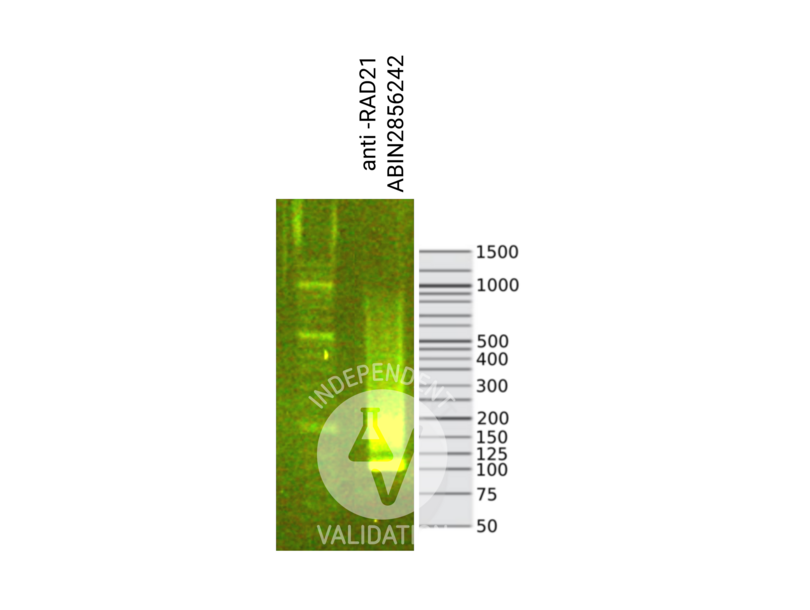
![Library profiles comparing fragment size distributions on an E-Gel EX 2% agarose gel (Thermo Fisher). Fragments obtained from CUT&RUN using RAD21 ABIN2856242 (right) after library preparation, compared to the E-Gel Sizing DNA Ladder (Thermo Fisher) (left).]() Library profiles comparing fragment size distributions on an E-Gel EX 2% agarose gel (Thermo Fisher). Fragments obtained from CUT&RUN using RAD21 ABIN2856242 (right) after library preparation, compared to the E-Gel Sizing DNA Ladder (Thermo Fisher) (left).
Library profiles comparing fragment size distributions on an E-Gel EX 2% agarose gel (Thermo Fisher). Fragments obtained from CUT&RUN using RAD21 ABIN2856242 (right) after library preparation, compared to the E-Gel Sizing DNA Ladder (Thermo Fisher) (left).
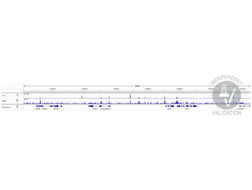
![1. Alignment tracks from CTCF CUT&RUN in HEK293T cells. 2. Alignment tracks from CUT&RUN targeting RAD21 in HEK293T cells using ABIN2856242 antibody showing the FBRS locus. 3. RefSeq Genes.]() 1. Alignment tracks from CTCF CUT&RUN in HEK293T cells. 2. Alignment tracks from CUT&RUN targeting RAD21 in HEK293T cells using ABIN2856242 antibody showing the FBRS locus. 3. RefSeq Genes.
Protocole
1. Alignment tracks from CTCF CUT&RUN in HEK293T cells. 2. Alignment tracks from CUT&RUN targeting RAD21 in HEK293T cells using ABIN2856242 antibody showing the FBRS locus. 3. RefSeq Genes.
Protocole -
-
Format
- Liquid
-
Concentration
- 1 mg/mL
-
Buffer
- 1XPBS, 20 % Glycerol ( pH 7). 0.025 % ProClin 300 was added as a preservative.
-
Agent conservateur
- ProClin
-
Précaution d'utilisation
- This product contains ProClin: a POISONOUS AND HAZARDOUS SUBSTANCE which should be handled by trained staff only.
-
Stock
- -20 °C
-
Stockage commentaire
- Keep as concentrated solution. Aliquot and store at -20°C or below. Avoid multiple freeze-thaw cycles.
-
-
-
: "Wnt signaling activation induces CTCF binding and loop formation at cis-regulatory elements of target genes." dans: Genome research, Vol. 35, Issue 8, pp. 1701-1716, (2025) (PubMed).
: "Genome-wide kinetic properties of transcriptional bursting in mouse embryonic stem cells." dans: Science advances, Vol. 6, Issue 25, pp. eaaz6699, (2020) (PubMed).
-
: "Wnt signaling activation induces CTCF binding and loop formation at cis-regulatory elements of target genes." dans: Genome research, Vol. 35, Issue 8, pp. 1701-1716, (2025) (PubMed).
-
- RAD21 (RAD21 Homolog (RAD21))
-
Autre désignation
- RAD21
-
Sujet
-
The protein encoded by this gene is highly similar to the gene product of Schizosaccharomyces pombe rad21, a gene involved in the repair of DNA double-strand breaks, as well as in chromatid cohesion during mitosis. This protein is a nuclear phospho-protein, which becomes hyperphosphorylated in cell cycle M phase. The highly regulated association of this protein with mitotic chromatin specifically at the centromere region suggests its role in sister chromatid cohesion in mitotic cells.
Cellular Localization: Nucleus -
Poids moléculaire
- 72 kDa
-
ID gène
- 5885
-
Pathways
- Positive Regulation of Endopeptidase Activity
Antigène
-


