CD63 anticorps (AA 120-175)

 (10 références)
 (3 validations)
Aperçu rapide pour CD63 anticorps (AA 120-175) (ABIN1440014)
Antigène
Voir toutes CD63 AnticorpsReactivité
Hôte
Clonalité
Conjugué
Application
-
-
Épitope
- AA 120-175
-
Fonction
- Anti-CD63
-
Séquence
- MENYPKNNHT ASILDRMQAD FKCCGAANYT DWEKIPSMSK NRVPDSCCI
-
Specificité
- Reacts with CD63, a 40-60 kDa glycoprotein, detected by Western blot in the following human (HeLa, HUH, Jurkat), mouse (AtT-20, Hepa, 3T3, RAW264.7), canine (MDCK) and monkey (COS-7) whole cell lysates.
-
Attributs du produit
- Lysosome-associated membrane glycoprotein 3
-
Purification
- Epitope affinity purified
-
Immunogène
- Purified recombinant peptide derived from within residues 120 aa to 175 aa of human CD63 produced in E. coli.
-
Isotype
- IgG
-
Informations sur le produit
-
À quoi peut servir l'anticorps CD63 ABIN1440014 ? Cet anticorps polyclonal de chèvre anti-CD63 n'est pas conjugué et convient pour détecter CD63, une glycoprotéine de 40-60 kDa une grande variété d'espèces (humain, souris, rat, chien et plus). L'anticorps CD63 est validé pour le FACS, l'IF, l'IHC et le Western blotting.
Quelles sont les données de validation disponibles pour cet anticorps CD63 ? 18 images pour une variété d'applications sont disponibles. L'anticorps est cité dans 10 publications PubMed et il dispose de 3 validations indépendantes de clients. Utilisez notre anticorps CD63 de haute qualité pour détecter de manière fiable le CD63.
Quelle est la fonction du CD63 ? Le CD63 fonctionne comme un récepteur de surface cellulaire pour le TIMP1 et joue un rôle dans l'activation des cascades de signalisation cellulaire. Joue un rôle dans l'activation de la signalisation de l'ITGB1 et des intégrines, conduisant à l'activation des kinases AKT, FAK/PTK2 et MAP. Favorise la survie cellulaire, la réorganisation du cytosquelette d'actine, l'adhésion cellulaire, l'étalement et la migration, via son rôle dans l'activation de AKT et FAK/PTK2. Joue un rôle dans la signalisation du VEGFA via son rôle dans la régulation de l'internalisation de KDR/VEGFR2. (UniProt)
-
-
anti-CD63 (CD63) antibody (Biotin)
CD63 Reactivité: Humain FACS, IHC (p), IP, ICC Hôte: Souris Monoclonal MEM-259 Biotin
anti-CD63 (CD63) antibody (APC)CD63 Reactivité: Humain FACS Hôte: Souris Monoclonal MEM-259 APC
anti-CD63 (CD63) antibodyCD63 Reactivité: Humain FACS, IHC (p), IP, ICC Hôte: Souris Monoclonal MEM-259 unconjugated
anti-CD63 (CD63) (AA 163-190), (C-Term) antibodyCD63 Reactivité: Humain FACS, WB, IHC (p) Hôte: Lapin Polyclonal RB26813 unconjugated
anti-CD63 (CD63) antibodyCD63 Reactivité: Humain FACS, WB, IHC, IF Hôte: Souris Monoclonal MX-49-129-5 unconjugated
anti-CD63 (CD63) (AA 103-203) antibodyCD63 Reactivité: Humain FACS, WB, IHC, IF, ELISA Hôte: Souris Monoclonal 1H1E11 unconjugated
anti-CD63 (CD63) (AA 103-203) antibodyCD63 Reactivité: Humain FACS, WB, IHC, IF, ELISA Hôte: Souris Monoclonal 10F11E6 unconjugated
anti-CD63 (CD63) antibody (PerCP)CD63 Reactivité: Humain FACS Hôte: Souris Monoclonal MEM-259 PerCP
anti-CD63 (CD63) antibody (PE)CD63 Reactivité: Humain FACS Hôte: Souris Monoclonal MEM-259 PE
anti-CD63 (CD63) (AA 103-203) antibodyCD63 Reactivité: Humain FACS, IHC, ELISA Hôte: Souris Monoclonal 1E8D4 unconjugated
-
-
Indications d'application
- WB:1:500-1:5,000, IF:1:25-1:250, IHC-P:1:250-1:1,000, IHC-F:1:250-1:1,000
-
Restrictions
- For Research Use only
-
-
- by
- Center for Diabetes and Metabolic Diseases, Indiana University
- No.
- #100043
- Date
- 30.08.2016
- Antigène
- CD63 antibody (CD63 Molecule) (AA 120-175)
- Numéro du lot
- 47220715
- Application validée
- Western Blotting
- Contrôle positif
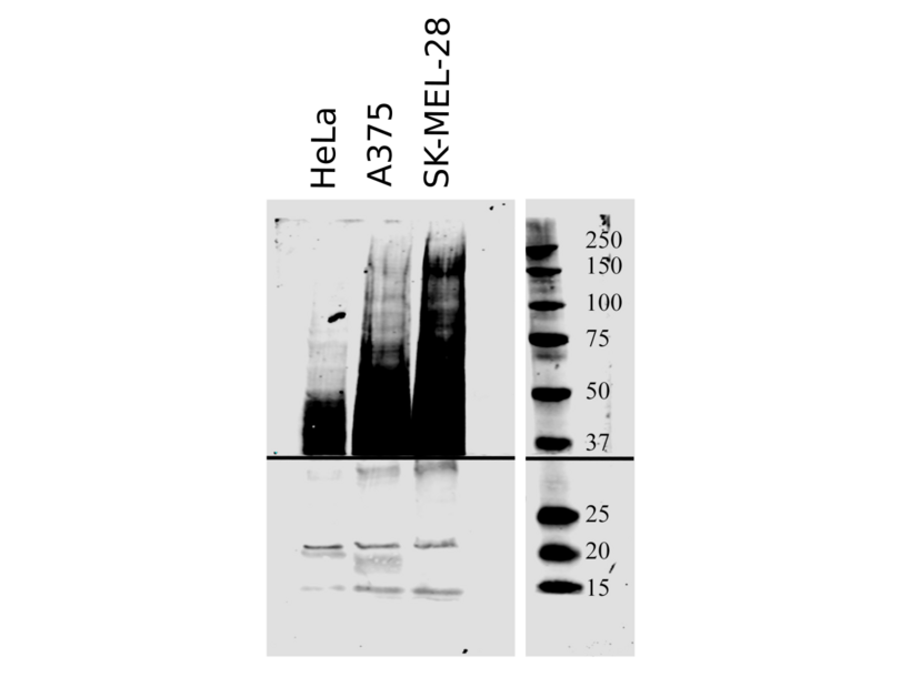
- A375 and SKMEL 28 melanoma cell line lysates
- Contrôle négative
- Conclusion
- The CD63 antibody ABIN1440014 works successfully in Western blot on human A375 and SKMEL 28 cell lysates.
- Anticorps primaire
- ABIN1440014
- Anticorps secondaire
- Donkey anti-Goat IgG (H+L) IRDye680RD (LiCor, antibodies-online product number ABIN2169630 , lot number: C50330-03)
- Full Protocol
- Cultured HeLa cells and human melanoma cell lines SK-MEL-28 and A375 were rinsed with PBS and lysed in 6M Urea/20mM Tris.
- 20µg protein from each samples were used for Western blot analysis.
- Samples were denatured in Bio-Rad Laemmli sample buffer (product 1610737, lot t350001755) containing beta-marcaptoethanol and separated on 4-20% Bio-Rad TGX gel (456-1034, lot t64041496).
- Transfer onto PVDF membrane (Millipore, product IPLF00010, lot R5EA5898E) was done on TransBlot SD apparatus (Bio-Rad) following the manufacturer’s protocol.
- Block membranes with LiCor Blocking Buffer (product 927-40000, lot V1381) for 2h.
- Incubation with primary CD63 antibody ABIN1440014 diluted 1:500 in LiCor Blocking Buffer at 4°C overnight.
- Wash 4x 15min with PBS-T.
- Incubation with secondary antibody LiCor Donkey anti-Goat IgG (Heavy & Light Chain) Antibody (IRDye680RD) secondary antibody (antibodies-online product number ABIN2169630, lot C50330-03, 1:15000 dilution) and incubated on a shaker for 1h at RT.
- Three rinses, followed by four 15min washes—2 PBS-T, 2 PBS (as required by LiCor).
- Blot was developed on a LiCor imaging system (Odyssey 9120, scan resolution: 169um, image quality: low).
- Notes
- The size marker was developed with a different fluorophore and is superimposed on the blot image for illustrative purposes. Melanoma lysates, those from A375 and SKMEL 28 cells served as positive controls for general cell lysates. Multiple bands with MW of 40-60kDa are expected to be revealed by ABIN1440014 in the cell lysates.
Validation #100043 (Western Blotting)
 Validation Images
![20µg total protein of cultured HeLa cells and human melanoma cell lines SKMEL28 and A375 were separated on an 4-20% polyacrylamid gel under denaturing conditions. Bands were revealed as described in the protocol sections with ABIN1440014 diluted 1:500.]() 20µg total protein of cultured HeLa cells and human melanoma cell lines SKMEL28 and A375 were separated on an 4-20% polyacrylamid gel under denaturing conditions. Bands were revealed as described in the protocol sections with ABIN1440014 diluted 1:500.
Protocole
20µg total protein of cultured HeLa cells and human melanoma cell lines SKMEL28 and A375 were separated on an 4-20% polyacrylamid gel under denaturing conditions. Bands were revealed as described in the protocol sections with ABIN1440014 diluted 1:500.
Protocole - by
- Center for Diabetes and Metabolic Diseases, Indiana University
- No.
- #100044
- Date
- 30.08.2016
- Antigène
- CD63 antibody (CD63 Molecule) (AA 120-175)
- Numéro du lot
- 47220715
- Application validée
- Western Blotting
- Contrôle positif
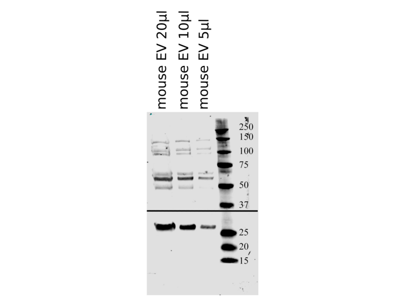
- C57BL/6 mouse serum
- Contrôle négative
- Conclusion
- Anticorps primaire
- ABIN1440014
- Anticorps secondaire
- Donkey anti-Goat IgG (H+L) IRDye680RD (LiCor, antibodies-online product number ABIN2169630 , lot number: C50330-03)
- Full Protocol
- Mouse serum was collected from C57BL/6 mice and lysed in 6M Urea/20mM Tris.
- Exosomes were extracted with ExoQuick reagent (SBI, product #EXOQ5A, lot #140624-001) following the manufacturer’s protocol and resuspended in 30µl of modified RIPA buffer.
- 20µl, 10µl, and 5µl aliquots of 5-fold diluted exosome solution were used.
- Samples were denatured in Bio-Rad Laemmli sample buffer (product # 1610737, lot #t350001755)containing beta-marcaptoethanol and separated on 4-20% Bio-Rad TGX gel (product #456-1034, lot #t64041496).
- Transfer onto PVDF membrane (Millipore, product #IPLF00010, lot #R5EA5898E) was done on TransBlot SD apparatus (Bio-Rad) following manufacturer’s protocol.
- Block membranes with LiCor Blocking Buffer (product 927-40000, lot V1381) for 2h.
- Incubation with
- primary CD63 antibody ABIN1440014 at 4°C diluted 1:500 in LiCor Blocking Buffer at 4°C overnight.
- primary rabbit anti-CD9 antibody (Proteintech, 20597-1-AP, lot 00013334) diluted 1:500 in LiCor Blocking Buffer at 4°C overnight.
- Wash 4x 15min with PBS-T.
- Incubation with secondary antibody LiCor Donkey anti-Goat IgG (Heavy & Light Chain) Antibody (IRDye680RD) secondary antibody (antibodies-online product number ABIN2169630, lot C50330-03, 1:15000 dilution) and incubated on a shaker for 1h at RT.
- Three rinses plus four 15min washes—2 PBS-T, 2 PBS (as required by LiCor).
- Blot was developed on a LiCor imaging system (Odyssey 9120, scan resolution: 169um, image quality: low).
- Notes
- C57BL/6 mouse serum served as positive control for extracellular vesicle (EV) samples. A single band around 55kDA is expected in EV samples.
- An alternative marker was used to confirm presence of EVs in the sample. Since CD63 is an exosomal marker, a quantitative correlation between the CD63 and the alternative marker signal strength with the amount of lysate loaded onto the gel is to be expected.
- The size marker was developed with a different fluorophore and is superimposed on the blot image for illustrative purposes.
- by
- Internal Disease Clinic, Faculty of Veterinary Medicine, University of Zagreb
- No.
- #101202
- Date
- 03.05.2017
- Antigène
- CD63
- Numéro du lot
- 47080912
- Application validée
- Western Blotting
- Contrôle positif
- extracellular vesicles from bovine follicular fluid, verified with Flotillin-1 antibody
- Contrôle négative
- non-enriched fractions
- Conclusion
- Passed. The CD63 antibody ABIN1440014 specifically reveals a faint band of the expected molecular weight in fractions enriched for exosomes.
- Anticorps primaire
- ABIN1440014
- Anticorps secondaire
- anti-goat-HRP
- Full Protocol
- Isolate extracellular vesicles from 0.5ml follicular fluid from cow obtained from abattoir using the qEV size-exclusion chromatography kit (iZON Science, qEVoriginal Size Exclusion Columns, NC0888135):
- Equilibrate columns with 10ml PBS.
- Apply the follicular fluid.
- Retain the first 3ml of the eluate as void volume negative control (V0).
- Collect the next three 0.5ml fractions (1, 2, 3) which are expected to contain extracellular vesicles.
- Collect next 0.5ml fraction (4) which is expected to contain proteins but not extracellular vesicles from follicular fluid.
- Concentrate fractions 25x using Microcon filters (Millipore, Ultracel YM-30, MRCF0R30) with a 25kDa cut-off.
- Boil all five fractions and 1µl of untreated follicular fluid as positive control in 1x Laemmli SDS sample buffer for 5min.
- Separate all protein in each fraction, 10µg for fraction 2, the richest in protein on a freshly cast 4% acrylamide stacking gel and 10% acrylamide separation SDS-PAGE gel.
- Transfer proteins onto a nitrocellulose membrane (GE Healthcare,Amersham Protran 0.45µm, 10600002) in an electroblotter tank (Biostep, electroblotting module GV100- EBGRM) Western blotting system using 20% methanol-containing transfer buffer for 2h at 150mA at RT.
- Block the membrane with Odyssey blocking buffer (PBS) (Odyssey, 927-400000) for 1h at RT.
- Incubation with primary goat anti-flotillin 1 antibody (antibodies-online, ABIN374222) diluted 1:500 in blocking buffer ON at 4°C.
- Wash membrane 3x 5min with TBST (TBS, 0.1% Tween-20).
- Incubate with secondary anti-goat-HRP antibody diluted 1:5000 in blocking buffer for 1h at RT.
- Wash membrane 3x 5min with TBST.
- Incubate membrane in Luminol for (Santa Cruz Biotechnology Inc., ImmunoCruz western blotting luminol reagent, SC-2048) 5min at RT.
- Reveal chimiuminescent bands using a chemiluminescence detector (LI-COR, Odyssey Fc, OFC-0966) imaging system.
- Strip membranes usingmild stripping protocol:
- Incubate membrane with stripping buffer (1.5% (w/v) glycine, 0.0% (w/v) SDS, 1% (v/v) tween 20, pH2.2) shaking for 2x 10min at RT.
- Wash membrane 2x 10min with PBS.
- Wash membrane2x 5min in TBS.
- Block the membrane with blocking buffer.
- Incubate with primary goat anti-CD63 antibody (antibodies-online, ABIN1440014, lot 0047080912) diluted 1:200 in blocking buffer ON at 4°C.
- Repeat the same steps as for the goat anti-flotillin 1 antibody.
- Notes
- We observed an albumin band in western blot of some fractions when we used a different secondary (HRP conjugated mouse anti-CD9 antibody) (not shown). Albumin was not observed with ABIN1440014.
- A problem we are going to solve by loading higher amount of sample and increasing primary antibody concentration is the weak band observed for CD63 due to the low abundance of the protein in this kind of exosomes.
-
-
Concentration
- 3 mg/mL
-
Buffer
- PBS, 20 % glycerol and 0.05 % sodium azide.
-
Agent conservateur
- Sodium azide
-
Précaution d'utilisation
- This product contains Sodium azide: a POISONOUS AND HAZARDOUS SUBSTANCE which should be handled by trained staff only.
-
Conseil sur la manipulation
- The antibody solution should be gently mixed before use.
-
Stock
- 4 °C,-20 °C
-
Stockage commentaire
- For continuous use, store at 2-8 C for one-two days. For extended storage, store in -20 C freezer. Working dilution samples should be discarded if not used within 12 hours.
-
-
-
: "Mass Spectrometry-Based Proteome Profiling of Extracellular Vesicles Derived from the Cerebrospinal Fluid of Adult Rhesus Monkeys Exposed to Cocaine throughout Gestation." dans: , Vol. 12, Issue 4, (2022) (PubMed).
: "Free fatty acids promote degranulation of azurophil granules in neutrophils by inducing production of NADPH oxidase-derived reactive oxygen species in cows with subclinical ketosis." dans: Journal of dairy science, Vol. 105, Issue 3, pp. 2473-2486, (2022) (PubMed).
: "Effects of general anaesthesia with an inhalational anaesthetic agent on the expression of exosomes in rats." dans: International journal of medical sciences, Vol. 19, Issue 9, pp. 1399-1407, (2022) (PubMed).
: "Investigation of canine extracellular vesicles in diffuse large B-cell lymphomas." dans: PloS one, Vol. 17, Issue 9, pp. e0274261, (2022) (PubMed).
: "Cardiosphere-derived exosomal microRNAs for myocardial repair in pediatric dilated cardiomyopathy." dans: Science translational medicine, Vol. 12, Issue 573, (2021) (PubMed).
: "Exosomes and STUB1/CHIP cooperate to maintain intracellular proteostasis." dans: PLoS ONE, Vol. 14, Issue 10, pp. e0223790, (2020) (PubMed).
: "Changes in localization and density of CD63-positive exosome-like substances in the hen oviduct with artificial insemination and their effect on sperm viability." dans: Theriogenology, Vol. 101, pp. 135-143, (2018) (PubMed).
: "Beta cell extracellular vesicle miR-21-5p cargo is increased in response to inflammatory cytokines and serves as a biomarker of type 1 diabetes." dans: Diabetologia, Vol. 61, Issue 5, pp. 1124-1134, (2018) (PubMed).
: "Exploration of serum- and cell culture-derived exosomes from dogs." dans: BMC veterinary research, Vol. 14, Issue 1, pp. 179, (2018) (PubMed).
: "Presence of Cx43 in extracellular vesicles reduces the cardiotoxicity of the anti-tumour therapeutic approach with doxorubicin." dans: Journal of extracellular vesicles, Vol. 5, pp. 32538, (2016) (PubMed).
-
-
- CD63
-
Autre désignation
- CD63
-
Sujet
-
CD63 is a member of the transmembrane 4 superfamily, also known as the tetraspanin family. Most of the members are cell-surface proteins that are characterized by the presence of four hydrophobic domains. These proteins mediate signal transduction events that play a role in the regulation of cell development, activation, growth and motility. This protein is a cell surface glycoprotein that is known to complex with integrins. It may function as a blood platelet activation marker.
CD63 antigen, CD63 antigen (melanoma 1 antigen), granulophysin, LAMP-3, lysosomal-associated membrane protein 3, lysosome-associated membrane glycoprotein 3, ME491, melanoma-associated antigen, melanoma 1 antigen, melanoma-associated antigen ME491, MLA1, ocular melanoma-associated antigen, OMA81H, tetraspanin-30, TSPAN30 antibody.
Antigène
-

 20µl, 10µl, and 5µl of 1:5 diluted extracellular vesicle (EV)/exosome solution from C57BL/6 mice were separated on an 4-20% polyacrylamid gel under denaturing conditions. CD63 (upper portion of the image) and CD9 (lower portion of the image) bands were revealed as described in the protocol section.
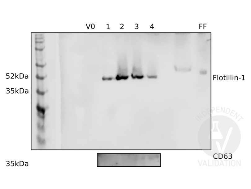 Different fractions from an enrichment of extracellular vesicles from bovine follicular fluid were probed with antibodies ABIN374222 and ABIN1440014 against exosome markers flotillin-1 (upper panel) and CD63 (lower panel). See protocol for more information. V0: void volume. 1: fraction 1, some exosomes expected. 2: fraction 2, usually the richest in exosomes. 3: third fraction, lower exosome concentration. 4: fourth fraction, contains no or very few exosomes. FF: follicular fluid.


