DVL1 anticorps (AA 21-100)
 (1 reference)
 (1 validation)
Aperçu rapide pour DVL1 anticorps (AA 21-100) (ABIN670671)
Antigène
Voir toutes DVL1 AnticorpsReactivité
Hôte
Clonalité
Conjugué
Application
-
-
Épitope
- AA 21-100
-
Réactivité croisée
- Humain, Souris, Rat
-
Purification
- Purified by Protein A.
-
Immunogène
- KLH conjugated synthetic peptide derived from human DVL1
-
Isotype
- IgG
-
-
anti-Dishevelled Segment Polarity Protein 1 (DVL1) (AA 442-470) antibody
DVL1 Reactivité: Humain, Souris WB, IF, IHC (p) Hôte: Lapin Polyclonal RB31716 unconjugated
anti-Dishevelled Segment Polarity Protein 1 (DVL1) (AA 150-285) antibodyDVL1 Reactivité: Humain WB, IHC Hôte: Lapin Polyclonal unconjugated
anti-Dishevelled Segment Polarity Protein 1 (DVL1) (AA 20-32) antibodyVerified DVL1 Reactivité: Humain IHC, ELISA Hôte: Chèvre Polyclonal unconjugated
anti-Dishevelled Segment Polarity Protein 1 (DVL1) (AA 510-640) antibodyDVL1 Reactivité: Humain WB, IHC Hôte: Lapin Polyclonal unconjugated
anti-Dishevelled Segment Polarity Protein 1 (DVL1) (Internal Region) antibodyVerified DVL1 Reactivité: Humain, Souris WB, IHC, ELISA Hôte: Chèvre Polyclonal unconjugated
anti-Dishevelled Segment Polarity Protein 1 (DVL1) (AA 1-110) antibodyDVL1 Reactivité: Humain WB, ELISA Hôte: Souris Polyclonal unconjugated
anti-Dishevelled Segment Polarity Protein 1 (DVL1) (AA 1-444) antibodyDVL1 Reactivité: Humain WB, PLA Hôte: Lapin Polyclonal unconjugated
anti-Dishevelled Segment Polarity Protein 1 (DVL1) (AA 1-444) antibodyDVL1 Reactivité: Humain WB Hôte: Souris Polyclonal unconjugated
anti-Dishevelled Segment Polarity Protein 1 (DVL1) (AA 70-120) antibodyDVL1 Reactivité: Humain, Souris, Rat, Singe WB Hôte: Lapin Polyclonal unconjugated
anti-Dishevelled Segment Polarity Protein 1 (DVL1) (AA 569-618) antibodyDVL1 Reactivité: Humain, Souris, Rat, Boeuf (Vache), Chien, Cheval WB Hôte: Lapin Polyclonal unconjugated
-
-
Indications d'application
-
WB 1:300-5000
ELISA 1:500-1000
FCM 1:20-100
IHC-P 1:200-400
IHC-F 1:100-500
IF(IHC-P) 1:50-200
IF(IHC-F) 1:50-200
IF(ICC) 1:50-200 -
Restrictions
- For Research Use only
-
-
- by
- Immunohistochemistry Core, NYU Langone
- No.
- #029657
- Date
- 03.04.2014
- Antigène
- Numéro du lot
- 140106
- Application validée
- Immunohistochemistry
- Contrôle positif
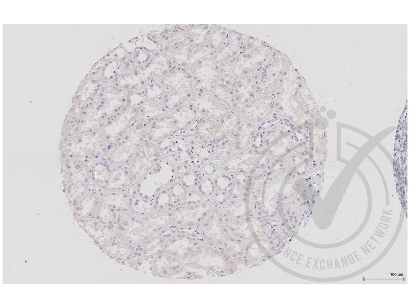
- Human kidney tubules
- Contrôle négative
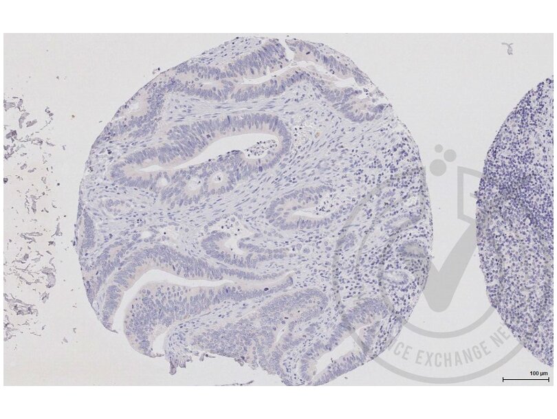
- Human colon stromal tissue
- Conclusion
- While faint, signal is detectable in positive control tissue and not in negative control tissue. Note that staining is expected in colon epithelial cells, and is not expected in kidney glomeruli.
- Anticorps primaire
- Antigen: Dishevelled, Dsh Homolog 1 (Drosophila) (DVL1)
- Catalog number: ABIN670671
- Lot number: 140106
- Anticorps secondaire
- Antibody: Biotinylated goat anti-rabbit/anti-mouse (Kit)
- Lot number: D07640BA
- Full Protocol
- Immunohistochemistry was performed on a Ventana NEXes automated platform; instrument manufacturer specific reagents are italicized.
- 1. Slides were preheated in convection oven at 60°C for 30 min
- 2. Deparaffinization procedure: - 3 changes of Xylene, 5 min each - 3 changes of 100% Ethanol, 3 min each - 3 changes of 95% Ethanol, 3 min each - Rinsed in distilled water, 3 changes
- 3. Heat retrieval procedure - Slides retrieved in 10.0 mM Citrate, pH6.0 in a 1000W microwave oven (~100°C) for 15 min. - Slides were allowed to cool (in citrate) for 30 min. - Slides were washed x 3 in Distilled water
- 4. NEXes instrument procedure, iView DAB paraffin protocol (*abridged*): - Slide chamber warmed to 37°C
- 5. Slides rinsed with *reaction buffer* x3
- 6. *iView Inhibitor (H2O2)* applied and incubated for 4 min
- 7. Slides rinsed with *reaction buffer*
- 8. Antibody Application - Primary antibody diluted 1:250 in PBS (100 microliter applied/slide) - Ventana Isotype control applied neat - Slides Incubated overnight at room temperature (~12 hours ~25°C)
- 9. Slides rinsed with *reaction buffer* x3
- 10. *iView Biotinylated IgG* applied and incubated for 8 min
- 11. Slides rinsed with *reaction buffer*
- 14. *iView Streptavidin-Horseradish Peroxidase* applied and incubated for 8 min
- 15. Slides rinsed with *reaction buffer*
- 16. *iView DAB/H2O2* applied and incubated for 8 min
- 17. Slides rinsed with *reaction buffer*
- 18. *iView Copper* applied and incubated for 4 min
- 19. Slides rinsed with *reaction buffer*
- 20. Slides washed in Dawn Detergent/tap water
- 21. Counterstain Procedure - Hematoxylin (Leica 560 MX) 30 sec - Slides washed in tap water, 1 min - Decolorized (10% Acetic Acid in 70% ethanol), 1 min - Slides washed in tap water, 1 min - Bluing (Austin Clear Ammonia), 1 min - Slides washed in tap water, 1 min
- 22. Dehydration/coverslipping procedure: - 3 changes of 95% Ethanol, 3 min each - 3 changes of 100% Ethanol, 3 min each - 3 changes of Xylene, 5 min each - Mounted with Permount
- 23. Imaging: Leica SCN 400F Whole Slide Scanner with Digital Image Hub and Leica Slidepath software
- Notes
- Step 1: Heated tissue 60°C for 30 minutes; manufacturer heats for 45 minutes.
- Step 2: No ethanol wash was performed during deparaffinization; manufacturer includes 1 wash of 80% ethanol for 3 minutes.
- Step 3.1: Slides were heated for 15 minutes; manufacturer provides a range of 15-20 minutes.
- Step 3.2: Slides were cooled for 30 minutes; manufacturer cools for 20 minutes.
- Step 4: Italicized reagents and incubation time are fixed instrument parameters.
- Step 5: Secondary species-specific serum block not used; manufacturer blocks with 5% normal goat serum for 2 hours.
- Step 8.1: Antibody diluted in PBS at 1:250; manufacture did not recommend diluent or dilution.
- Step 8.2.1: Primary antibody incubated at room temperature overnight; manufacturer incubates overnight 4°C with agitation.
Validation #029657 (Immunohistochemistry)
 Validation Images
![Figure 1: Human kidney tissue stained with anti-DVL1 (brown) and counterstained with hematoxylin.]() Figure 1: Human kidney tissue stained with anti-DVL1 (brown) and counterstained with hematoxylin.
Figure 1: Human kidney tissue stained with anti-DVL1 (brown) and counterstained with hematoxylin.
![Figure 2: Human colon tissue stained with anti-DVL1 (brown) and counterstained with hematoxylin.]() Figure 2: Human colon tissue stained with anti-DVL1 (brown) and counterstained with hematoxylin.
Figure 2: Human colon tissue stained with anti-DVL1 (brown) and counterstained with hematoxylin.
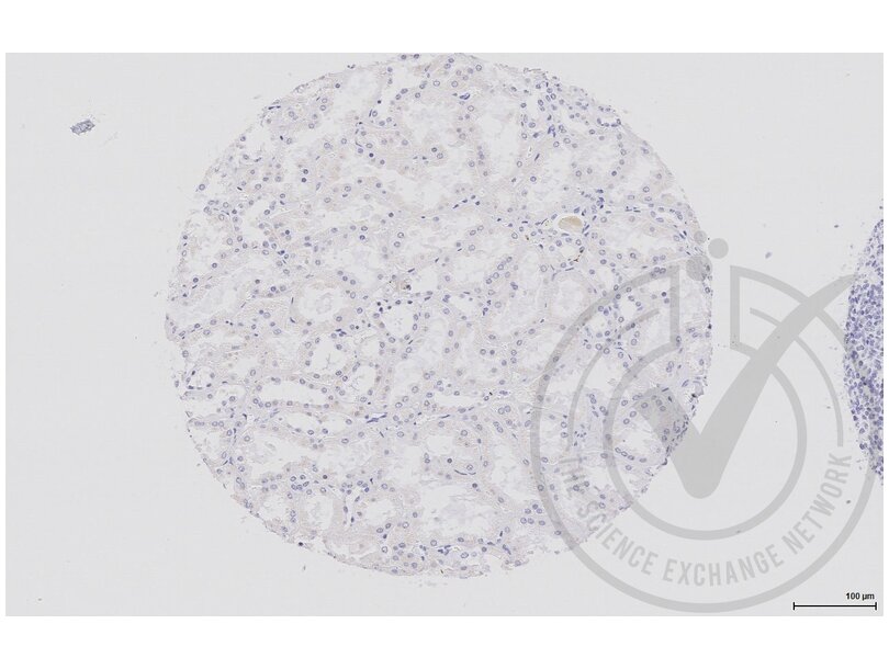
![Figure 3: Human kidney tissue stained with isotype control antibody (brown) and counterstained with hematoxylin.]() Figure 3: Human kidney tissue stained with isotype control antibody (brown) and counterstained with hematoxylin.
Figure 3: Human kidney tissue stained with isotype control antibody (brown) and counterstained with hematoxylin.
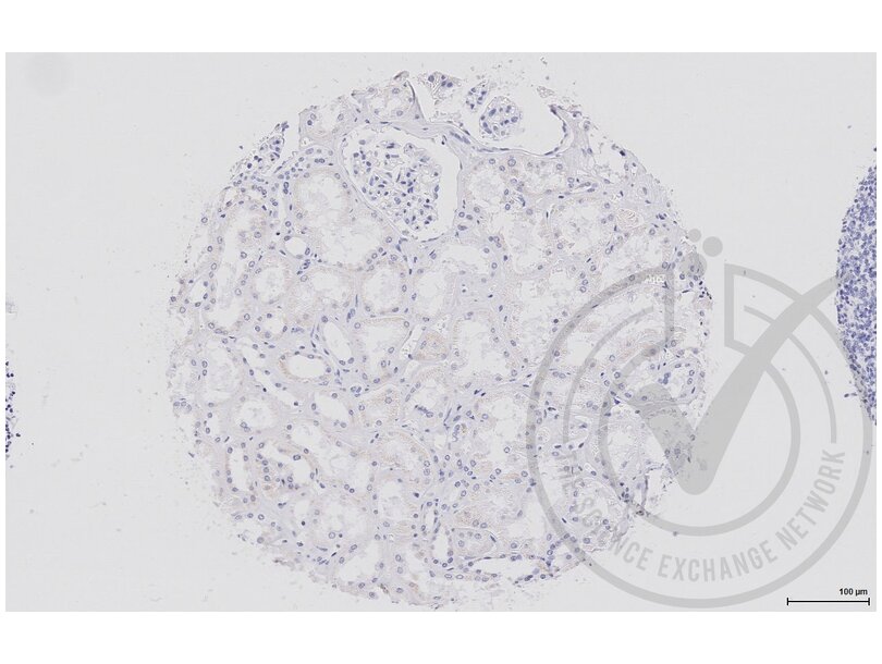
![Figure 4: Human kidney tissue stained with secondary only (brown) and counterstained with hematoxylin.]() Figure 4: Human kidney tissue stained with secondary only (brown) and counterstained with hematoxylin.
Protocole
Figure 4: Human kidney tissue stained with secondary only (brown) and counterstained with hematoxylin.
Protocole -
-
Format
- Liquid
-
Concentration
- 1 μg/μL
-
Buffer
- 0.01M TBS( pH 7.4) with 1 % BSA, 0.02 % Proclin300 and 50 % Glycerol.
-
Agent conservateur
- ProClin
-
Précaution d'utilisation
- This product contains ProClin: a POISONOUS AND HAZARDOUS SUBSTANCE, which should be handled by trained staff only.
-
Stock
- 4 °C,-20 °C
-
Stockage commentaire
- Shipped at 4°C. Store at -20°C for one year. Avoid repeated freeze/thaw cycles.
-
Date de péremption
- 12 months
-
-
-
: "Wnt4/?-catenin signaling pathway modulates balloon-injured carotid artery restenosis via disheveled-1." dans: International journal of clinical and experimental pathology, Vol. 7, Issue 12, pp. 8421-31, (2015) (PubMed).
-
: "Wnt4/?-catenin signaling pathway modulates balloon-injured carotid artery restenosis via disheveled-1." dans: International journal of clinical and experimental pathology, Vol. 7, Issue 12, pp. 8421-31, (2015) (PubMed).
-
- DVL1 (Dishevelled Segment Polarity Protein 1 (DVL1))
-
Autre désignation
- DVL1
-
Sujet
-
Synonyms: DVL, DVL1L1, DVL1P1, Segment polarity protein dishevelled homolog DVL-1, Dishevelled-1, DSH homolog 1, DVL1
Background: Participates in Wnt signaling by binding to the cytoplasmic C-terminus of frizzled family members and transducing the Wnt signal to down-stream effectors. Plays a role both in canonical and non-canonical Wnt signaling. Plays a role in the signal transduction pathways mediated by multiple Wnt genes. Required for LEF1 activation upon WNT1 and WNT3A signaling. DVL1 and PAK1 form a ternary complex with MUSK which is important for MUSK-dependent regulation of AChR clustering during the formation of the neuromuscular junction (NMJ).
-
ID gène
- 1855
-
UniProt
- O14640
-
Pathways
- Signalisation WNT, Synaptic Membrane, Skeletal Muscle Fiber Development
Antigène
-


