NOTCH2 anticorps
 (3 références)
 (1 validation)
Aperçu rapide pour NOTCH2 anticorps (ABIN104899)
Antigène
Voir toutes NOTCH2 AnticorpsReactivité
Hôte
Clonalité
Conjugué
Application
-
-
N° du produit (Fournisseur)
- 100-401-408
-
Fournisseur
- Rockland
-
Fonction
- NOTCH 2 Antibody
-
Réactivité croisée (Details)
- This antiserum is directed against human NOTCH2. The immunogen epitope is only exposed after gamma secretase cleavage and is not accessible in the uncleaved form.
-
Attributs du produit
- Synonyms: rabbit anti-Notch2 antibody, AGS2 antibody, hN2 antibody, Notch homolog 2 antibody, neurogenic locus notch homolog protein 2
-
Purification
- Antiserum
-
Stérilité
- Sterile filtered
-
Immunogène
-
Immunogen: This whole rabbit serum was prepared by repeated immunizations with a synthetic peptide corresponding to amino acid residues of human Notch 2 located near the N-terminal sequence of the cleaved N intracellular domain (NICD).
Immunogen Type: Conjugated Peptide
-
Informations sur le produit
-
A quoi peut servir l'anticorps NOTCH2 ABIN104899 ? Cet anticorps polyclonal non conjugué de lapin anti-NOTCH2 détecte de manière fiable le NOTCH2 humain par ELISA et Western blotting.
Quelles sont les données de validation disponibles pour cet anticorps NOTCH2 ? Pour le Western blotting, une validation indépendante est actuellement disponible et a été réalisée à l'Université d'Ulm, en Allemagne. Sur la base de la séquence, on peut supposer que l'anticorps NOTCH2 fonctionne également pour les espèces de rat et de souris. L'anticorps a été utilisé dans trois publications à ce jour, qui sont indiquées ci-dessous et peuvent être lues sur PubMed. Il y a 2 images disponibles qui démontrent la performance de l'anticorps NOTCH2 en Western blot. Utilisez notre anticorps NOTCH2 pour détecter de manière fiable le NOTCH2 humain.
Quelle est la fonction de NOTCH2 ? NOTCH2 fonctionne comme un récepteur pour les ligands membranaires Jagged-1 (JAG1), Jagged-2 (JAG2) et Delta-1 (DLL1) afin de réguler la détermination du statut cellulaire. Lors de l'activation du ligand par le domaine intracellulaire de l'encoche (NICD) libéré, il forme un complexe activateur de transcription avec RBPJ/RBPSUH et active les gènes du locus enhancer of split. Affecte la mise en œuvre des programmes de différenciation, de prolifération et d'apoptose (Par similarité). NOTCH2 semble être impliqué dans le remodelage et l'homéostasie osseuse. En collaboration avec RELA/p65 augmente l'activité du promoteur de NFATc1 et régule positivement la différenciation des ostéoclastes induite par RANKL. (UniProt)
-
-
anti-Notch 2 (NOTCH2) (Asp1733), (cleaved) antibody
NOTCH2 Reactivité: Humain, Souris, Rat WB, ELISA, IHC Hôte: Lapin Polyclonal unconjugated
anti-Notch 2 (NOTCH2) (C-Term) antibodyNOTCH2 Reactivité: Humain, Souris WB, ChIP-seq Hôte: Lapin Polyclonal unconjugated
anti-Notch 2 (NOTCH2) (cleaved), (Internal Region) antibodyNOTCH2 Reactivité: Humain WB, ELISA, IHC Hôte: Lapin Polyclonal unconjugated
anti-Notch 2 (NOTCH2) (AA 2242-2471) antibodyNOTCH2 Reactivité: Humain WB, IF Hôte: Lapin Polyclonal unconjugated
anti-Notch 2 (NOTCH2) (AA 59-250) antibodyNOTCH2 Reactivité: Humain ELISA, IHC, IF Hôte: Lapin Polyclonal unconjugated
anti-Notch 2 (NOTCH2) (AA 2251-2466) antibodyNOTCH2 Reactivité: Humain WB, IHC, IP, ICC Hôte: Lapin Polyclonal unconjugated
anti-Notch 2 (NOTCH2) (AA 1501-1700) antibodyNOTCH2 Reactivité: Humain, Souris WB, ELISA, IF (cc), IF (p), IHC (p), IHC (fro) Hôte: Lapin Polyclonal unconjugated
anti-Notch 2 (NOTCH2) (cleaved), (Internal Region) antibodyNOTCH2 Reactivité: Humain WB, ELISA Hôte: Lapin Polyclonal unconjugated
anti-Notch 2 (NOTCH2) (Cleaved-Ala1734), (Internal Region) antibodyNOTCH2 Reactivité: Humain, Souris, Rat WB, ELISA Hôte: Lapin Polyclonal unconjugated
-
-
Indications d'application
-
Immunohistochemistry Dilution: 1:500
Application Note: Anti-Notch2 has been tested in ELISA, WB, and IHC. Anti-NOTCH-2 has a strong response was detected by ELISA against the immunizing peptide. This product was assayed against the peptide immunogen in a standard capture ELISA using Peroxidase conjugated anti-Rabbit IgG [H&L] (Goat) (code #611-1302) and ABTS (2,2'-azino-bis-[3-ethylbenthiazoline-6-sulfonic acid]) (code # ABTS-100) as a substrate for 30 minutes at room temperature. A working dilution of 1:30,000 to 1:90,000 is suggested in ELISA this product.
Western Blot Dilution: 1:400 - 1:2,000
ELISA Dilution: 1:20,000 - 1:100,000
Other: User Optimized
-
Restrictions
- For Research Use only
-
-
- by
- AG Pancreatic Development and Stem cell differentiation, Universitätsklinikum Ulm
- No.
- #103703
- Date
- 16.06.2019
- Antigène
- NOTCH2
- Numéro du lot
- Application validée
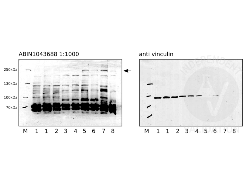
- Western Blotting
- Contrôle positif
pancreatic endoderm cells (day 10)
pancreatic progenitor cells (day 14)
- Contrôle négative
human pluripotent stem cells (day 0)
defnite endoderm cells (day 4)
- Conclusion
ABIN1043688 reveals two protein bands of the expected size and some weaker extraneous bands in cell lysates of human pluripotent stem cells, definite endoderm cells, pancreatic endoderm cells, and pancreatic progenitor cells.
- Anticorps primaire
- ABIN1043688
- Anticorps secondaire
- donkey anti-rabbit HRP-conjugated antibody (GE Healthcare, NA9310V)
- Full Protocol
- Grow HUES8 in mTeSR1 (STEMCELL Technologies, 85850) at 37°C and 5% O2, 5% CO2 in 2ml in a 6-well plate to 90% confluency.
- Harvest cells using TrypLE (ThermoFisher Scientific, 12604013) following the manufacturer´s instructions.
- Lyse 2x10⁶ cells in 30µl per well cold RIPA buffer (50mM Tris-HCl pH 8.0, 150mM NaCl, 0.1% SDS, 0.5% deoxycholate, 1% TritonX 100 in ddH2O) supplemented with 1mM PMSF and 1x protease inhibitor (Roche, 11836170001) for 30min on ice with 3x vortexing in 1.5ml microcentrifuge tubes.
- Centrifuge tubes at 10000xg for 8min at 4°C.
- Transfer supernatant to a new 1.5ml microcentrifuge tube and store at -80°C.
- Determine total protein content of the lysates using a Bradford assay (Bio-Rad, 500-0006).
- Denature 50µg of total protein for 5min at 95°C in 30µl 1x Laemmli SDS sample buffer and subsequently separate them on a denaturing 7.5% polyacrylamide gel (7.5% Acrylamide, 0.375M Tris-HCl pH8.8, 0.1% SDS, 0.1% APS, 0.1% TEMED) for about 120min at 120V.
- Transfer proteins onto a PVDF membrane (Sigma Aldrich, IPVH00010) with transfer buffer (5.27g Tris, 2.93 g glycerine, 200 ml methanol, fill to 1l ddH2O) in a semidry western blotting system for 80min at 80mA/gel.
- Check transfer of the separated proteins by Ponceau S staining.
- Rinse membrane with water.
- Wash membrane for 5min with TBST (TBS, 0.02% Tween20).
- Block the membrane with 20ml blocking buffer (TBST, 5% milk) on a shaker for 1h at RT.
- Rinse membrane 3x with TBST.
- Wash membrane on a shaker 3x for 5min with TBST.
- Shrink-wrap and incubate membrane with primary
- rabbit anti-NOTCH2 antibody (antibodies-online, ABIN1043688) diluted 1:1000 respectively in blocking buffer ON at 4°C or
- mouse anti-vinculin antibody (SigmaAldrich, V9264) diluted 1:1000 in blocking buffer at RT for 1h.
- Wash membrane 3x for 5min with TBST.
- Incubate membrane with secondary
- donkey anti-rabbit HRP-conjugated antibody (GE Healthcare, NA9310V) diluted 1:5000 in TBST containing 1% milk for 1h at RT or
- donkey anti-mouse HRP-conjugated antibody (GE Healthcare, NA9340V) diluted 1:5000 in TBST containing 1% milk for 1h at RT.
- Wash membrane 3x for 5min with TBST.
- Reveal protein bands using ECL solution (ThermoScientific, 34076) with an exposure time of 40min for the ABIN1043688 and 2min for the vinculin loading control.
- Notes
ABIN1043688 revealed some strong unspecific bands. Nevertheless, a band over 250kDa appears to be highly specific, as it is only detected at day 10 (lanes 5 and 6) and day 14 (lanes 7 and 8).
Manual exposure had to be performed due to a defect of the fusion detector in our institute.
ABIN1043688 was also tested in IHC on 4-5µm FFPE sections of primary human kidney cancer tissue. Epitope retrieval was carried out using Tris-EDTA buffer at pH9.0 (Zytomed, ZUC029-500), EDTA at pH8.0 (Leica, RE7116), or citrate buffer at pH6.1 (Agilent, S169984-2) for 20min in a decloaking chamber.
Interpretation of the results was difficult, as the method of antigen retrieval strongly effects the staining pattern. Antigen retrieval using the citrate buffer at pH6 might be ok, but knockout or knockdown studies are necessary to clearly conclusively show specificity.
Validation #103703 (Western Blotting)
 Validation Images
![Western blot with ABIN1043688 on human pluripotent stem cells (1 and 2), definite endoderm cells (3 and 4), pancreatic endoderm cells (5 and 6), and pancreatic progenitors (7 and 8). ABIN1043688 was used at a 1:1000 dilution. Vinculin served as loading control.]() Western blot with ABIN1043688 on human pluripotent stem cells (1 and 2), definite endoderm cells (3 and 4), pancreatic endoderm cells (5 and 6), and pancreatic progenitors (7 and 8). ABIN1043688 was used at a 1:1000 dilution. Vinculin served as loading control.
Protocole
Western blot with ABIN1043688 on human pluripotent stem cells (1 and 2), definite endoderm cells (3 and 4), pancreatic endoderm cells (5 and 6), and pancreatic progenitors (7 and 8). ABIN1043688 was used at a 1:1000 dilution. Vinculin served as loading control.
Protocole -
-
Format
- Liquid
-
Concentration
- 70 mg/mL
-
Buffer
-
Buffer: 0.02 M Potassium Phosphate, 0.15 M Sodium Chloride, pH 7.2
Stabilizer: None
Preservative: 0.01 % (w/v) Sodium Azide -
Agent conservateur
- Sodium azide
-
Précaution d'utilisation
- This product contains Sodium azide: a POISONOUS AND HAZARDOUS SUBSTANCE which should be handled by trained staff only.
-
Stock
- 4 °C,-20 °C
-
Stockage commentaire
- Store vial at -20° C prior to opening. Aliquot contents and freeze at -20° C or below for extended storage. Avoid cycles of freezing and thawing. Centrifuge product if not completely clear after standing at room temperature. This product is stable for several weeks at 4° C as an undiluted liquid. Dilute only prior to immediate use.
-
Date de péremption
- 12 months
-
-
-
: "Potassium depletion induces cellular conversion in the outer medullary collecting duct altering Notch signaling pathway." dans: Scientific reports, Vol. 10, Issue 1, pp. 5708, (2020) (PubMed).
: "Notch signaling and inherited disease syndromes." dans: Human molecular genetics, Vol. 12 Spec No 1, pp. R9-13, (2003) (PubMed).
: "Notch-1 and Notch-2 exhibit unique patterns of expression in human B-lineage cells." dans: Leukemia, Vol. 14, Issue 12, pp. 2095-102, (2001) (PubMed).
-
: "Potassium depletion induces cellular conversion in the outer medullary collecting duct altering Notch signaling pathway." dans: Scientific reports, Vol. 10, Issue 1, pp. 5708, (2020) (PubMed).
-
- NOTCH2 (Notch 2 (NOTCH2))
-
Autre désignation
- NOTCH2
-
Sujet
- Background: Anti-Notch 2 Antibody recognizes Notch2 that is synthesized in the endoplasmic reticulum as an inactive form which is proteolytically cleaved by a furin-like convertase (S1 cleavage) in the trans-golgi network before it reaches the plasma membrane to yield an active, ligand-accessible form. Cleavage results in a C-terminal fragment N(TM) and a N-terminal fragment N(EC). Following ligand binding, it is cleaved (S2 cleavage) by TNF-alpha converting enzyme (TACE) to yield a membrane-associated intermediate fragment called Notch extracellular truncation (NEXT). This fragment is then cleaved by presenilin-dependent gamma-secretase (S3 cleavage) to release the intracellular domain (NICD) from the membrane. Anti-NOTCH2 Antibody is useful for researchers interested in Notch1, Jagged1, Jagged2, and Delta1, as well as neuroscience, transcription and cancer research.
-
ID gène
- 4853, 24041035
-
UniProt
- Q04721
-
Pathways
- Signalisation Notch, Stem Cell Maintenance
Antigène
-


