IDE anticorps (AA 491-590)
 (1 validation)
Aperçu rapide pour IDE anticorps (AA 491-590) (ABIN723665)
Antigène
Voir toutes IDE AnticorpsReactivité
Hôte
Clonalité
Conjugué
Application
-
-
Épitope
- AA 491-590
-
Réactivité croisée
- Souris
-
Homologie
- Human,Rat,Cow,Pig,Chicken
-
Purification
- Purified by Protein A.
-
Immunogène
- KLH conjugated synthetic peptide derived from human IDE
-
Isotype
- IgG
-
-
anti-Insulin-Degrading Enzyme (IDE) (AA 491-590) antibody
IDE Reactivité: Humain, Souris, Rat WB, ELISA, IHC (p), IF (cc), IF (p), IHC (fro) Hôte: Lapin Polyclonal unconjugated
anti-Insulin-Degrading Enzyme (IDE) (AA 753-973) antibodyIDE Reactivité: Humain WB, IHC Hôte: Lapin Polyclonal unconjugated
anti-Insulin-Degrading Enzyme (IDE) (AA 1-250) antibodyIDE Reactivité: Humain WB, IF Hôte: Lapin Polyclonal unconjugated
anti-Insulin-Degrading Enzyme (IDE) antibodyIDE Reactivité: Humain WB, IF, IHC (p) Hôte: Souris Monoclonal 1H4 unconjugated
anti-Insulin-Degrading Enzyme (IDE) (Internal Region) antibodyVerified IDE Reactivité: Humain WB, IHC, ELISA Hôte: Chèvre Polyclonal unconjugated
anti-Insulin-Degrading Enzyme (IDE) (AA 920-1019) antibodyIDE Reactivité: Humain WB, ELISA Hôte: Souris Polyclonal unconjugated
anti-Insulin-Degrading Enzyme (IDE) (N-Term) antibodyIDE Reactivité: Humain, Souris, Rat WB, IHC, ELISA Hôte: Lapin Polyclonal unconjugated
anti-Insulin-Degrading Enzyme (IDE) antibodyKD Validated IDE Reactivité: Humain WB, FACS Hôte: Lapin Monoclonal 23GB6210 unconjugated Recombinant Antibody
anti-Insulin-Degrading Enzyme (IDE) (AA 501-800) antibodyIDE Reactivité: Humain IHC, ELISA, IF Hôte: Lapin Polyclonal unconjugated
anti-Insulin-Degrading Enzyme (IDE) antibodyIDE Reactivité: Humain WB Hôte: Souris Monoclonal 3H4 unconjugated
-
-
Indications d'application
-
WB 1:300-5000
ELISA 1:500-1000
IHC-P 1:200-400
IHC-F 1:100-500 -
Restrictions
- For Research Use only
-
-
- by
- Prof. Merighi, Laboratory of Neurobiology, Department of Veterinary Sciences, University of Turin
- No.
- #104436
- Date
- 15.03.2023
- Antigène
- IDE
- Numéro du lot
- 9C07M588
- Application validée
- Immunohistochemistry
- Contrôle positif
Adult mouse liver and brain fixed in 4% paraformaldehyde
- Contrôle négative
One control slice for each experimental procedure processed omitting the primary antibody; overnight incubation in diluent solution only.
- Conclusion
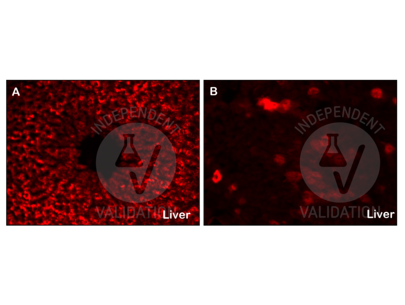
Passed. The IDE antibody (AA 491-590) ABIN723665 works in IHC-P at 1:50, 1:100 and 1:300 concentrations with tyramide amplification.
- Anticorps primaire
- ABIN723665
- Anticorps secondaire
- poly-HRP conjugated goat anti-rabbit antibody
- Full Protocol
- Perfuse mice with paraformaldehyde 4% in 0.1 M phosphate buffer pH 7.4 and post-fix in the same fixative for an additional 2 h at RT.
- Wash, dehydrate, and embed samples in paraffin wax.
- Wash several times with 0.01 M PBS.
- Cut liver with a microtome into 20 µm sections and mount on glass slides.
- After paraffin removal, incubate sections for 1 h at RT in PBS containing 1% albumin from chicken egg white (Sigma, A5378) and 0.3% Triton-X-100 (BioRad, 161-0407, lot 00583) to block non-specific binding sites.
- Incubate sections with primary rabbit anti-IDE (antibodies-online, ABIN723665, lot BB03153570) diluted 1:50, 1:100, 1:200, 1:300, and 1:400 in PBS-BSA-PLL ON at RT in a humid chamber.
- Wash sections 3x 5 min with 0.01 M PBS.
- Incubate sections with secondary poly-HRP conjugated goat anti-rabbit antibody from Alexa Fluor 594 Tyramide SuperBoost Kit, goat anti-rabbit IgG (Thermo Fisher Scientific, B40915, lot 2086865) for 1 h at RT.
- Wash sections 3x 5 min with 0.01 M PBS.
- I• Incubate sections with Tyramide working solution containing 100X Tyramide stock solution (Alexa 594), 100X H2O2 solution and 1X Reaction buffer for 10 min.
- Stop the reaction with the Reaction Stop Reagent working solution.
- Wash sections 3x 5 min with 0.01M PBS.
- Mount specimens in Fluoroshield (Sigma, F6182, lot MKCB0153V).
- Acquire images Leica DM 6000B fluorescence microscope equipped with a digital camera at 40x magnification.
- Notes
Validation #104436 (Immunohistochemistry)
 Validation Images
![Staining with ABIN723665 of IDE-positive hepatocytes in the adult mouse liver. A: low magnification view of a hepatic lobule CV = central vein. B = high magnification of a group of IDE-stained hepatocytes]() Staining with ABIN723665 of IDE-positive hepatocytes in the adult mouse liver. A: low magnification view of a hepatic lobule CV = central vein. B = high magnification of a group of IDE-stained hepatocytes
Staining with ABIN723665 of IDE-positive hepatocytes in the adult mouse liver. A: low magnification view of a hepatic lobule CV = central vein. B = high magnification of a group of IDE-stained hepatocytes
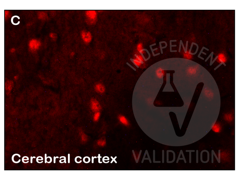
![Staining with ABIN723665 of IDE-positive neurons in adult mouse brain.]() Staining with ABIN723665 of IDE-positive neurons in adult mouse brain.
Protocole
Staining with ABIN723665 of IDE-positive neurons in adult mouse brain.
Protocole -
-
Format
- Liquid
-
Concentration
- 1 μg/μL
-
Buffer
- 0.01M TBS( pH 7.4) with 1 % BSA, 0.02 % Proclin300 and 50 % Glycerol.
-
Agent conservateur
- ProClin
-
Précaution d'utilisation
- This product contains ProClin: a POISONOUS AND HAZARDOUS SUBSTANCE, which should be handled by trained staff only.
-
Stock
- 4 °C,-20 °C
-
Stockage commentaire
- Shipped at 4°C. Store at -20°C for one year. Avoid repeated freeze/thaw cycles.
-
Date de péremption
- 12 months
-
-
- IDE (Insulin-Degrading Enzyme (IDE))
-
Autre désignation
- IDE
-
Sujet
-
Synonyms: BC2, Insulin degrading enzyme, FLJ35968, insulin protease, insulinase, insulysin, Abeta-degrading protease, FLJ35968, Ide, IDE_HUMAN, Insulin-degrading enzyme, OTTHUMP00000020097.
Background: Insulysin was identified nearly a century ago as an enzyme responsible for the degradation of insulin in cells, although the precise interactions between insulin and insulysin remain elusive. Human insulysin was cloned in 1988, and shown to be a 118 kDa protein that exists primarily as a homodimer, and perhaps also complexed with other molecules. The sequence is well conserved between humans, rats and mice, and the antibody recognizes these species. Insulysin is a metalloproteinase of the clan ME, family M16, which contains an active site HxxEH, a reversal of the canonical HExxH zinc binding motif. Considered a zinc metalloproteinase, the activity of insulysin can be blocked with EDTA or 1-10 phenanthroline. In addition to the active metalloproteinase domain, insulysin contains a second metalloproteinase site which is considered catalytically inactive, and is thought to assist in substrate binding. Insulysin is most closely related to the bacterial proteinase pitrilysin, (the human orthologue of which appears to be MPRP1) and the mammalian proteinsae nardilysin. Generally thought to be a cytoplasmic protein, insulysin has been isolated from many different tissues and cell lines, and can degrade intact insulin, insulin B chain, glucagon, denatured hemoglobin, alpha amyloid protein, TGF alpha and amylin. Recent work implicates insulysin in clearing beta amyloid plaques from the brain, and has generated much interest in Alzheimer?s disease research. The pH optimum for insulysin is basic, pH 8.5, which also distinguishes it from other metalloproteinases. Insulin degrading enzyme (IDE) has a preferential affinity for insulin such that the presence of insulin will inhibit IDE mediated degradation of other substrates. IDE degrades a variety of other peptides including atrial natriuretic peptide and amylin.
-
ID gène
- 3416
-
Pathways
- SARS-CoV-2 Protein Interactome
Antigène
-


