IDE anticorps (AA 491-590)
-
- Antigène Voir toutes IDE Anticorps
- IDE (Insulin-Degrading Enzyme (IDE))
-
Épitope
- AA 491-590
-
Reactivité
- Humain, Souris, Rat
-
Hôte
- Lapin
-
Clonalité
- Polyclonal
-
Conjugué
- Cet anticorp IDE est non-conjugé
-
Application
- Western Blotting (WB), ELISA, Immunohistochemistry (Paraffin-embedded Sections) (IHC (p)), Immunofluorescence (Cultured Cells) (IF (cc)), Immunofluorescence (Paraffin-embedded Sections) (IF (p)), Immunohistochemistry (Frozen Sections) (IHC (fro))
- Réactivité croisée
- Humain, Souris, Rat
- Homologie
- Cow,Pig,Chicken
- Purification
- Purified by Protein A.
- Immunogène
- KLH conjugated synthetic peptide derived from human IDE
- Isotype
- IgG
-
-
- Indications d'application
-
WB 1:300-5000
ELISA 1:500-1000
IHC-P 1:200-400
IHC-F 1:100-500
IF(IHC-P) 1:50-200
IF(IHC-F) 1:50-200
IF(ICC) 1:50-200 - Restrictions
- For Research Use only
-
- by
- Prof. Merighi, Laboratory of Neurobiology, Department of Veterinary Sciences, University of Turin
- No.
- #104435
- Date
- 15.03.2023
- Antigène
- IDE
- Numéro du lot
- 9C07M588
- Application validée
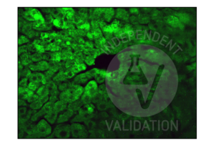
- Immunohistochemistry
- Contrôle positif
Adult mouse liver fixed in 4% paraformaldehyde
- Contrôle négative
One control slice for each experimental procedure processed omitting the primary antibody; overnight incubation in diluent solution only.
- Conclusion
Passed. The IDE antibody (AA 491-590) ABIN723680 works in IHC-P at 1:100 concentrations with Tyramide amplification.
- Anticorps primaire
- ABIN723680
- Anticorps secondaire
- poly-HRP conjugated goat anti-rabbit antibody
- Full Protocol
- Perfuse mice with paraformaldehyde 4% in 0.1 M phosphate buffer pH 7.4 and post-fix in the same fixative for an additional 2 h at RT.
- Wash, dehydrate, and embed samples in paraffin wax.
- Wash several times with 0.01 M PBS.
- Cut liver with a microtome into 20 µm sections and mount on glass slides.
- After paraffin removal, incubate sections for 1 h at RT in PBS containing 1% albumin from chicken egg white (Sigma, A5378) and 0.3% Triton-X-100 (BioRad, 161-0407, lot 00583) to block non-specific binding sites.
- Incubate sections with primary rabbit anti-IDE (antibodies-online, ABIN723680, lot 9C07M588) diluted 1:50, 1:100, 1:200, and 1:300 in PBS-BSA-PLL ON at RT in a humid chamber.
- Wash sections 3x 5 min with 0.01 M PBS.
- Incubate sections with secondary poly-HRP conjugated goat anti-rabbit antibody from Alexa Fluor 488 Tyramide SuperBoost Kit, goat anti-rabbit IgG (Thermo Fisher Scientific, B40922, lot 2465062) for 1 h at RT.
- Wash sections 3x 5 min with 0.01 M PBS.
- Incubate sections with Tyramide working solution containing 100X Tyramide stock solution (Alexa 488), 100X H2O2 solution and 1X Reaction buffer for 10 min.
- Stop the reaction with the Reaction Stop Reagent working solution.
- Wash sections 3x 5 min with 0.01M PBS.
- Mount specimens in Fluoroshield (Sigma, F6182, lot MKCB0153V).
- Acquire images with a fluorescence microscope and appropriate filter settings for AF488, e.g. Leica DM 6000B fluorescence microscope equipped with a digital camera at 40x magnification.
- Notes
Validation #104435 (Immunohistochemistry)![Testé avec succès 'Independent Validation' signe]()
![Testé avec succès 'Independent Validation' signe]() Validation ImagesProtocole
Validation ImagesProtocole -
- by
- Prof. Merighi, Laboratory of Neurobiology, Department of Veterinary Sciences, University of Turin
- No.
- #104497
- Date
- 15.03.2023
- Antigène
- IDE
- Numéro du lot
- 9C07M588
- Application validée
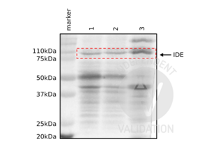
- Western Blotting
- Contrôle positif
Adult mouse brain, cerebellum, and liver
- Contrôle négative
- Conclusion
Passed. The IDE antibody (AA 491-590) ABIN723680 works in WB at 1:1000 concentrations with sensitive ECL substrate.
- Anticorps primaire
- ABIN723680
- Anticorps secondaire
- HRP-conjugated mouse anti-rabbit
- Full Protocol
- Homogenize tissues with cold lysis buffer containing 50 mM Tris HCl, 150 mM NaCl, 1% Triton X-100, 1 mM EDTA, and 1% protease inhibitor (Sigma P8340) using an ultrasonic homogenizer (MSE, SoniPrep 150) with 16 amplitude, 20 s on, 10 s off pulse for 60 s.
- Centrifuge tissue homogentates at 13,000 rpm for 20 min at 4 °C.
- Collect supernatants and Determine total protein content using a Bradford assay.
- Denature 50 µg of total protein for 5 min at 90 °C and subsequently separate them on a denaturing 12% PAGE-SDS gel alongside a Precision Plus Protein Dual Color Standard (Bio-Rad, 160374).
- Electro-transfer proteins onto nitrocellulose membrane (Amerscham Biosciences, RPN203D) ON in the cold room.
- Wash membrane 3x for 10 mon with 0.01 M PBS containing 0.1% Tween-20 (PBST).
- Block membrane with PBST containing 2% bovine serum albumin for 1 h at RT.
- Incubate membrane with primary rabbit anti-IDE antibody (antibodies-online, ABIN723680, lot 9C07M588) diluted 1:1,000 in PBST ON at 4 °C.
- Wash membrane 3x 10 min with PBST.
- Incubate membrane with secondary HRP-conjugated mouse anti-rabbit IgG (Sigma, A1949) diluted 1:4,000 in PBST for 1 h at RT.
- Wash membrane 3x 10 min with PBST.
- Visualize proteins with WesternBright Sirius HRP substrate (Advansta, K-12043) using a ChemiDoc Imaging System.
- Notes
Validation #104497 (Western Blotting)![Testé avec succès 'Independent Validation' signe]()
![Testé avec succès 'Independent Validation' signe]() Validation ImagesProtocole
Validation ImagesProtocole -
- Format
- Liquid
- Concentration
- 1 μg/μL
- Buffer
- 0.01M TBS( pH 7.4) with 1 % BSA, 0.02 % Proclin300 and 50 % Glycerol.
- Agent conservateur
- ProClin
- Précaution d'utilisation
- This product contains ProClin: a POISONOUS AND HAZARDOUS SUBSTANCE, which should be handled by trained staff only.
- Stock
- 4 °C,-20 °C
- Stockage commentaire
- Shipped at 4°C. Store at -20°C for one year. Avoid repeated freeze/thaw cycles.
- Date de péremption
- 12 months
-
- Antigène
- IDE (Insulin-Degrading Enzyme (IDE))
- Autre désignation
- IDE (IDE Produits)
- Synonymes
- anticorps zgc:162603, anticorps IDE, anticorps 1300012G03Rik, anticorps 4833415K22Rik, anticorps AA675336, anticorps AI507533, anticorps INSDEGM, anticorps INSULYSIN, anticorps insulin degrading enzyme, anticorps insulin-degrading enzyme, anticorps metallopeptidase, anticorps IDE, anticorps ide, anticorps LOC591315, anticorps CC1G_01115, anticorps Bm1_26615, anticorps PTRG_10875, anticorps SJAG_01430, anticorps EBI_21700, anticorps VDBG_06702, anticorps LOAG_08082, anticorps PGTG_04982, anticorps Tsp_00424, anticorps Ide
- Sujet
-
Synonyms: INSULYSIN, Insulin-degrading enzyme, Abeta-degrading protease, Insulin protease, Insulinase, IDE
Background: Plays a role in the cellular breakdown of insulin, IAPP, glucagon, bradykinin, kallidin and other peptides, and thereby plays a role in intercellular peptide signaling. Degrades amyloid formed by APP and IAPP. May play a role in the degradation and clearance of naturally secreted amyloid beta-protein by neurons and microglia.
- ID gène
- 3416
- UniProt
- P14735
- Pathways
- SARS-CoV-2 Protein Interactome
-


 (2 validations)


