GDNF anticorps (AA 121-211)
 (1 reference)
 (1 validation)
Aperçu rapide pour GDNF anticorps (AA 121-211) (ABIN736536)
Antigène
Voir toutes GDNF AnticorpsReactivité
Hôte
Clonalité
Conjugué
Application
-
-
Épitope
- AA 121-211
-
Réactivité croisée
- Boeuf (Vache), Humain, Souris, Lapin, Rat
-
Homologie
- Dog,Cow,Pig,Horse,Chicken
-
Purification
- Purified by Protein A.
-
Immunogène
- KLH conjugated synthetic peptide derived from human GDNF
-
Isotype
- IgG
-
-
anti-Glial Cell Line Derived Neurotrophic Factor (GDNF) antibody
GDNF Reactivité: Humain WB, IHC, IP, ELISA, FACS Hôte: Lapin Monoclonal 11D2 unconjugated Recombinant Antibody
anti-Glial Cell Line Derived Neurotrophic Factor (GDNF) (AA 142-158), (Middle Region) antibodyGDNF Reactivité: Humain, Rat WB, IHC Hôte: Lapin Polyclonal unconjugated
anti-Glial Cell Line Derived Neurotrophic Factor (GDNF) (AA 8-36) antibodyGDNF Reactivité: Humain, Souris WB, IHC, ELISA, FACS Hôte: Lapin Polyclonal unconjugated
anti-Glial Cell Line Derived Neurotrophic Factor (GDNF) (AA 192-207), (Mature) antibodyGDNF Reactivité: Humain WB, IHC, IF, IC Hôte: Lapin Polyclonal unconjugated
anti-Glial Cell Line Derived Neurotrophic Factor (GDNF) (AA 78-211) antibodyGDNF Reactivité: Humain WB, IHC, IP, ICC Hôte: Lapin Polyclonal unconjugated
anti-Glial Cell Line Derived Neurotrophic Factor (GDNF) (AA 78-211) antibodyGDNF Reactivité: Rat WB, IHC, IP, ICC Hôte: Lapin Polyclonal unconjugated
anti-Glial Cell Line Derived Neurotrophic Factor (GDNF) (AA 79-217) antibodyGDNF Reactivité: Souris WB, IHC, IP, ICC Hôte: Lapin Polyclonal unconjugated
anti-Glial Cell Line Derived Neurotrophic Factor (GDNF) antibodyGDNF Reactivité: Humain WB, FACS Hôte: Lapin Monoclonal 24GB3395 unconjugated Recombinant Antibody
anti-Glial Cell Line Derived Neurotrophic Factor (GDNF) (AA 8-36), (N-Term) antibodyGDNF Reactivité: Humain FACS, IHC (p) Hôte: Lapin Polyclonal RB21995 unconjugated
anti-Glial Cell Line Derived Neurotrophic Factor (GDNF) (AA 121-211) antibody (FITC)GDNF Reactivité: Humain, Souris, Rat, Boeuf (Vache), Lapin WB, FACS, IF (cc), IF (p) Hôte: Lapin Polyclonal FITC
-
-
Indications d'application
-
WB 1:300-5000
ELISA 1:500-1000
FCM 1:20-100
IHC-P 1:200-400
IHC-F 1:100-500
IF(IHC-P) 1:50-200
IF(IHC-F) 1:50-200
IF(ICC) 1:50-200 -
Restrictions
- For Research Use only
-
-
- by
- Prof. Merighi, Laboratory of Neurobiology, Department of Veterinary Sciences, University of Turin
- No.
- #102003
- Date
- 12.02.2018
- Antigène
- GDNF
- Numéro du lot
- 9F01M8
- Application validée
- Immunofluorescence
- Contrôle positif
- mouse hippocampus and putamen
- Contrôle négative
- no primary control
- Conclusion
ABIN736536 works in IF albeit with some background staining that we have been unable to eliminate although using different dilutions to increase the signal-to-noise ratio.
- Anticorps primaire
- ABIN736536
- Anticorps secondaire
- anti-rabbit AF488 conjugated antibody (Life Technologies)
- Full Protocol
- Perfuse mouse with 4% paraformaldehyde in 0.1M phosphate buffer (PB) pH7.4.
- Post-fix spinal cord and brain blocks in the same fixative for additional 2h at RT.
- Wash spinal cord and brain blocks several times with PBS.
- Cut blocks with a vibratome (Leica, VT1000 S) into 70µm thick transverse sections.
- Cut dorsal root ganglions (DRGs) with a cryostat into 17µm thick sections after cryoprotection and glass mounting.
- Block free floating vibratome and glass mounted cryostat sections with blocking solution (0.01M PBS5% Normal Goat Serum (NGS; Sigma, G9023, lot SLBV1396), 0.1% Triton X-100 (BioRad, 161-0407, lot 00583) for 1h at RT.
- Incubate sections with primary rabbit anti-GDNF antibody (antibodies-online, ABIN736536, lot 9F01M8) diluted 1:100 in blocking solution ON at RT.
- Wash sections 4x for 5min with 0.01M PBS.
- Incubate sections with secondary goat anti-rabbit AF488 conjugated antibody (Life Technologies) diluted 1:500 in PBS for 1h at RT.
- Wash sections 4x for 5min with 0.01M PBS.
- Mount sections in Fluoroshield (Sigma, F6182, lot MKCB0153V).
- Notes
Different dilutions were also tested (1:200, 1:500, 1:2000) with or without Triton-X but the 1:100 dilution and the use of Triton-X in the blocking solution gave the best results.
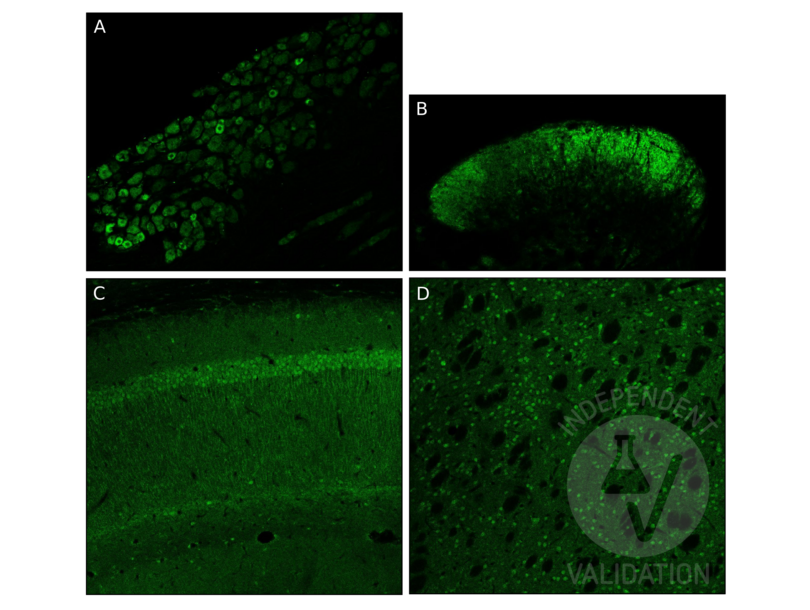
Staining is mainly present in the cytoplasm of small-size neurons in the DRG, consistent with what has been previously described. ABIN736536 stains fibers in superficial laminae of the dorsal horn of the spinal cord. In the hippocampus staining is present in the CA1 both in the pyramidal neurons (cell body and dendrites) and in individual interneurons. In the putamen ABIN736536 stains neuron cell bodies and some dendrites.
Validation #102003 (Immunofluorescence)
 Validation Images
![IF staining with ABIN736536 of mouse dorsal root ganglion (DRG, A), the dorsal horn of the spinal cord (B), and the hippocampus CA1 (C) and Putamen (D).]() IF staining with ABIN736536 of mouse dorsal root ganglion (DRG, A), the dorsal horn of the spinal cord (B), and the hippocampus CA1 (C) and Putamen (D).
Protocole
IF staining with ABIN736536 of mouse dorsal root ganglion (DRG, A), the dorsal horn of the spinal cord (B), and the hippocampus CA1 (C) and Putamen (D).
Protocole -
-
Format
- Liquid
-
Concentration
- 1 μg/μL
-
Buffer
- 0.01M TBS( pH 7.4) with 1 % BSA, 0.02 % Proclin300 and 50 % Glycerol.
-
Agent conservateur
- ProClin
-
Précaution d'utilisation
- This product contains ProClin: a POISONOUS AND HAZARDOUS SUBSTANCE, which should be handled by trained staff only.
-
Stock
- 4 °C,-20 °C
-
Stockage commentaire
- Shipped at 4°C. Store at -20°C for one year. Avoid repeated freeze/thaw cycles.
-
Date de péremption
- 12 months
-
-
-
: "Normalization of ventral tegmental area structure following acupuncture in a rat model of heroin relapse." dans: Neural regeneration research, Vol. 9, Issue 3, pp. 301-7, (2014) (PubMed).
-
: "Normalization of ventral tegmental area structure following acupuncture in a rat model of heroin relapse." dans: Neural regeneration research, Vol. 9, Issue 3, pp. 301-7, (2014) (PubMed).
-
- GDNF (Glial Cell Line Derived Neurotrophic Factor (GDNF))
-
Autre désignation
- GDNF
-
Sujet
-
Synonyms: ATF1, ATF2, HSCR3, HFB1-GDNF, Glial cell line-derived neurotrophic factor, hGDNF, Astrocyte-derived trophic factor, ATF, GDNF
Background: Neurotrophic factor that enhances survival and morphological differentiation of dopaminergic neurons and increases their high-affinity dopamine uptake.
-
ID gène
- 2668
-
UniProt
- P39905
-
Pathways
- Signalisation RTK, Synaptic Membrane, Tube Formation, Autophagy, Smooth Muscle Cell Migration
Antigène
-


