RDH11 anticorps
 (1 validation)
Aperçu rapide pour RDH11 anticorps (ABIN966957)
Antigène
Voir toutes RDH11 AnticorpsReactivité
Hôte
Clonalité
Conjugué
Application
-
-
anti-Retinol Dehydrogenase 11 (All-Trans/9-Cis/11-Cis) (RDH11) (AA 22-318) antibody
RDH11 Reactivité: Humain WB, ELISA, IHC Hôte: Lapin Polyclonal unconjugated
anti-Retinol Dehydrogenase 11 (All-Trans/9-Cis/11-Cis) (RDH11) (AA 24-318) antibodyRDH11 Reactivité: Humain WB, ELISA Hôte: Souris Monoclonal 1H6 unconjugated
anti-Retinol Dehydrogenase 11 (All-Trans/9-Cis/11-Cis) (RDH11) antibodyRDH11 Reactivité: Humain, Rat WB Hôte: Lapin Polyclonal unconjugated
anti-Retinol Dehydrogenase 11 (All-Trans/9-Cis/11-Cis) (RDH11) (C-Term) antibodyRDH11 Reactivité: Humain, Rat, Cobaye, Cheval, Porc, Lapin, Roussette (Chauve-souris), Singe WB Hôte: Lapin Polyclonal unconjugated
anti-Retinol Dehydrogenase 11 (All-Trans/9-Cis/11-Cis) (RDH11) (C-Term) antibodyRDH11 Reactivité: Humain, Souris, Rat, Cobaye, Cheval, Porc, Lapin, Boeuf (Vache), Chien WB Hôte: Lapin Polyclonal unconjugated
-
-
Restrictions
- For Research Use only
-
-
- by
- Palczewski Lab, Center For Translational Vision Research, UC Irvine
- No.
- #104470
- Date
- 23.03.2023
- Antigène
- RDH11
- Numéro du lot
- 0618
- Application validée
- Immunohistochemistry
- Contrôle positif
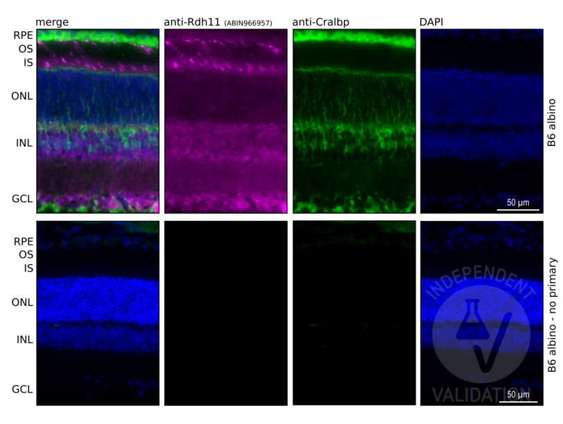
Retina cryosection from B6 Albino (B6(Cg)-Tyrc-2J/J) animal
- Contrôle négative
Retina cryosection from B6 Albino (B6(Cg)-Tyrc-2J/J) animal
No primary antibody
- Conclusion
Passed. Presence of specific signal in the RPE cell layer was considered as indication of specific immunoreactivity using the anti-RDH11 antibody ABIN966957.
- Anticorps primaire
- ABIN966957
- Anticorps secondaire
- donkey anti-rabbit AF647-conjugated antibody (Abcam, 150075)
- Full Protocol
- Collect eyes from mice and fix with paraformaldehyde 4% (Electron Microscopy Sciences, 15710) in 1x PBS for 30 min at RT.
- Cryoprotection with sucrose series:
- Wash in 10% sucrose in 1x PBS.
- Immerse in 10% sucrose in 1x PBS for 30 min at RT.
- Wash in 20% sucrose in 1x PBS.
- Immerse in 20% sucrose in 1x PBS for 30 min RT.
- Wash in 30% sucrose in 1x PBS.
- 30% sucrose ON at 4°C.
- Embed eyes in OCT compound (Tissue-Tek O.C.T. Compound, 4583).
- Cut retinal sections at a thickness of 12 μm on a cryostat.
- Air dry sections for 15 min at RT, store at -80°C until use.
- Bring sections to RT and rehydrate in 1x PBS for 1 h.
- Incubate sections in blocking buffer (1x PBS, 3% BSA (Sigma-Aldrich, A7030), 3% Donkey serum (Sigma-Aldrich, S30-100ML), 0.1% Triton X-100 (Sigma-Aldrich, X100-500ML)) for 1 h at RT.
- Incubate sections with primary rabbit anti-RDH11 antibody (antibodies-online, ABIN966957, lot 0618) diluted 1:50 in blocking buffer ON at RT. Include a no primary antibody negative controls. Additionally, counterstaing with primary mouse anti-CRALBP antibody (Thermo Fisher Scientific, MA1-813).
- Incubate sections with secondary AF647-conjugated donkey anti-rabbit antibody (Abcam, Ab150075) or AF488-conjugated donkey anti-mouse antibody (Thermo Fisher Scientific, A32766) diluted 1:500 in blocking buffer for 1 h at RT.
- Rinse sections once with 1x PBS, 0.1% Triton X-100 for 5 min at RT.
- Incubate sections in 1x DAPI (Thermo Fisher Scientific, 62248) in 1x PBS, 0.1% Triton X-100 for 15 min at RT.
- Rinse sections 3x with 1x PBS, 0.1% Triton X-100 for 5 min at RT.
- Mount sections in VECTASHIELD® HardSet™ Antifade Mounting Medium (Vector Laboratories, H-1400) mounting medium.
- Acquire images with a fluorescence microscope and appropriate filter settings.For the validation purposes Keyence BZ-X800E fluorescence microscope was used with following filters: BZ-X DAPI for DAPI, BZ-X GFP for AF488, BZ-X Cy5 for AF647. Images were taken at 10x and 40x magnification.
- Notes
Experiment involved validation of the specificity of 4 antibodies against different Rdh proteins: Rdh5 (ABIN7254060), Rdh10 (ABIN7118460), Rdh11 (ABIN966957), and Rdh12 (ABIN7167836). All 4 proteins are important for eye function and highly expressed in neural retina and/or RPE. Validation is based on comparison of each staining with known pattern of expression in the mouse retina. For review highlighting each Rdh localization see PMID20801113.
To aid orientation in the cell layers anti-Cralbp counterstain was included in the staining (Thermo MA1-813). Cralbp (Rlbp1) is highly expressed in RPE and Müller glia cells in mouse retina.
Validation #104470 (Immunohistochemistry)
 Validation Images
![Retinal sections from the wild-type (B6 albino) mice immunostained with anti-RDH11 antibody ABIN966957. DAPI staining shows localization of the inner (INL) and outer (ONL) nuclear layer of the mouse retina. Cralbp (Rlbp1) co-staining was used to visualize RPE and Müller glia cells in the retina. Presence of specific signal in the RPE cell layer confirms specific immunoreactivity.]() Retinal sections from the wild-type (B6 albino) mice immunostained with anti-RDH11 antibody ABIN966957. DAPI staining shows localization of the inner (INL) and outer (ONL) nuclear layer of the mouse retina. Cralbp (Rlbp1) co-staining was used to visualize RPE and Müller glia cells in the retina. Presence of specific signal in the RPE cell layer confirms specific immunoreactivity.
Protocole
Retinal sections from the wild-type (B6 albino) mice immunostained with anti-RDH11 antibody ABIN966957. DAPI staining shows localization of the inner (INL) and outer (ONL) nuclear layer of the mouse retina. Cralbp (Rlbp1) co-staining was used to visualize RPE and Müller glia cells in the retina. Presence of specific signal in the RPE cell layer confirms specific immunoreactivity.
Protocole -
-
Stock
- 4 °C
-
-
- RDH11 (Retinol Dehydrogenase 11 (All-Trans/9-Cis/11-Cis) (RDH11))
-
Autre désignation
- RDH11
Antigène
-


