LRP2 anticorps (AA 3401-3500) (Cy3)
 (1 validation)
-
- Antigène Voir toutes LRP2 Anticorps
- LRP2 (Low Density Lipoprotein Receptor-Related Protein 2 (LRP2))
-
Épitope
- AA 3401-3500
-
Reactivité
- Humain, Souris
-
Hôte
- Lapin
-
Clonalité
- Polyclonal
-
Conjugué
- Cet anticorp LRP2 est conjugé à/à la Cy3
-
Application
- Flow Cytometry (FACS), Immunofluorescence (Cultured Cells) (IF (cc)), Immunofluorescence (Paraffin-embedded Sections) (IF (p))
- Réactivité croisée
- Humain, Souris
- Homologie
- Rat,Dog,Horse,Chicken,Rabbit
- Purification
- Purified by Protein A.
- Immunogène
- KLH conjugated synthetic peptide derived from human Megalin
- Isotype
- IgG
-
anti-Low Density Lipoprotein Receptor-Related Protein 2 (LRP2) (AA 3401-3500) antibody
LRP2 Reactivité: Humain, Souris ELISA, FACS, IF (cc), IF (p), IHC (p), ICC, IHC (fro) Hôte: Lapin Polyclonal unconjugated
anti-Low Density Lipoprotein Receptor-Related Protein 2 (LRP2) (AA 202-332) antibodyLRP2 Reactivité: Humain ELISA, IHC, IF Hôte: Lapin Polyclonal unconjugated
anti-Low Density Lipoprotein Receptor-Related Protein 2 (LRP2) antibodyLRP2 Reactivité: Souris, Rat IHC, IF Hôte: Lapin Polyclonal unconjugated
anti-Low Density Lipoprotein Receptor-Related Protein 2 (LRP2) (AA 4035-4184) antibodyLRP2 Reactivité: Humain IHC, WB, IP, ICC Hôte: Lapin Polyclonal unconjugated
anti-Low Density Lipoprotein Receptor-Related Protein 2 (LRP2) (AA 4446-4655) antibodyLRP2 Reactivité: Humain IHC, WB Hôte: Lapin Polyclonal unconjugated
anti-Low Density Lipoprotein Receptor-Related Protein 2 (LRP2) (AA 1211-1369) antibodyLRP2 Reactivité: Humain ELISA, IHC Hôte: Lapin Polyclonal unconjugated
anti-Low Density Lipoprotein Receptor-Related Protein 2 (LRP2) (AA 4626-4655), (C-Term) antibodyLRP2 Reactivité: Humain IHC (p) Hôte: Lapin Polyclonal RB1834 unconjugated
anti-Low Density Lipoprotein Receptor-Related Protein 2 (LRP2) antibodyLRP2 Reactivité: Humain, Souris, Rat ELISA, IHC Hôte: Lapin Polyclonal unconjugated
anti-Low Density Lipoprotein Receptor-Related Protein 2 (LRP2) (Internal Region) antibodyLRP2 Reactivité: Humain, Souris, Rat ELISA, IHC, WB Hôte: Lapin Polyclonal unconjugated
-
- Indications d'application
-
FCM 1:20-100
IF(IHC-P) 1:50-200
IF(IHC-F) 1:50-200
IF(ICC) 1:50-200 - Restrictions
- For Research Use only
-
- by
- CaresBio Laboratory
- No.
- #029611
- Date
- 11.02.2014
- Antigène
- Numéro du lot
- YEYY9
- Application validée
- Immunofluorescence
- Contrôle positif
- MCF7 cells
- Contrôle négative
- HeLa cells
- Conclusion
- Strong signal was detected in positive control tissues and not in negative control tissues. Note that there was a small amount of signal generated in the negative control sample.
- Anticorps primaire
- Antigen: Low Density Lipoprotein Receptor-Related Protein 2 (LRP2) antibody (Cy3)
- Catalog number: ABIN750991
- Lot number: YEYY9
- Anticorps secondaire
- Used only for the secondary only control, as primary antibody was directly conjugated to Cy3
- Antibody: Cy3 Goat Anti-Rabbit IgG (H+L)
- Full Protocol
- MCF7 and HeLa cell lines were grown directly on coverslips and fixed with 4% formaldehyde in PBS for 15 min at room temperature (RT).
- Fixed cells were rinsed three times in PBS for 5 min each at RT.
- Cells were blocked in 1X PBS/1% BSA/0.3% Triton™ X-100 to block unspecific binding of the antibodies for 60 min at RT.
- Cells were incubated with fluorophore conjugated primary antibody diluted 1:250 and 1:100 in 1X PBS/1% BSA/0.3% Triton™ X-100 overnight at 4°C.
- Cells were rinsed three times in PBS for 5 min each at RT.
- Coverslips were mounted on slides with ProLong® Gold Antifade Reagent with DAPI.
- Isotype control staining:
- All the steps are done same as previously described until cells were blocked in 1X PBS/1% BSA/0.3% Triton™ X-100 to block unspecific binding of the antibodies for 60 min at RT. - Cells were incubated with 10% normal rabbit serum overnight at 4°C.
- Cells were rinsed three times in PBS for 5 min each at RT.
- Cells were incubated with goat anti rabbit CY3 conjugated secondary antibody for 60 min in dark at RT.
- Cells were rinsed three times in PBS for 5 min each at RT.
- Coverslips were mounted on slides with ProLong® Gold Antifade Reagent with DAPI. Secondary only staining:
- All the steps are done same as previously described until cells were blocked in 1X PBS/1% BSA/0.3% Triton™ X-100 overnight at 4°C. - Cells were incubated with goat anti rabbit CY3 conjugated secondary antibody for 60 min in dark at RT.
- Cells were rinsed three times in PBS for 5 min each at RT.
- Coverslips were mounted on slides with ProLong® Gold Antifade Reagent with DAPI.
- Stained cells were imaged with a Nikon C2+ confocal microscope.
- Notes
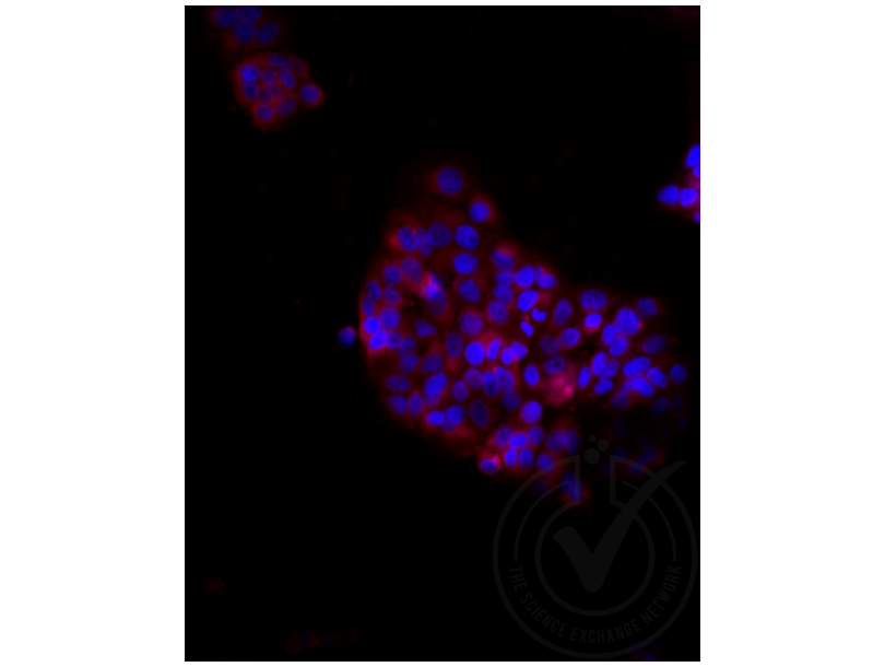
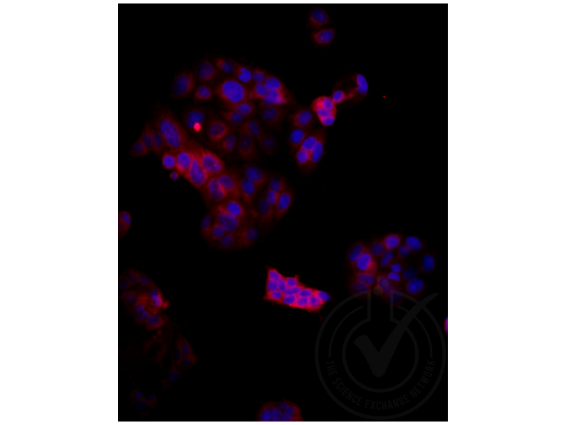
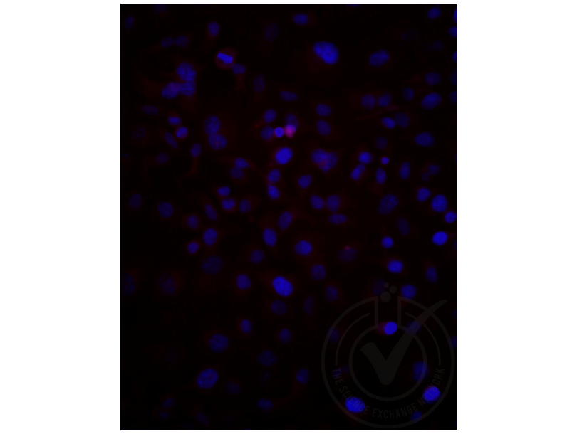
- We did the first set of experiment with 1:250 dilution of the primary antibody but have observed lower level of expression of LRP2 in MCF7 (positive) cells (Figure 2). We repeated experiment with higher concentration of the primary antibody (1:100) and the expression level increased as shown in Figure 1. We have noticed lower level of expression of LRP2 in HeLa (negative) cells (Figure 3). We have used rabbit normal serum for isotype control to match the species with primary antibody (rabbit) and also we used bovine albumin serum (BSA) as blocking and antibody diluent buffer to reduce species cross reactivity.
Validation #029611 (Immunofluorescence)
 Validation Images
![Figure 1: MCF7 cells stained at 1:250 with LRP2 (red) and a nuclear counterstain (blue).]() Figure 1: MCF7 cells stained at 1:250 with LRP2 (red) and a nuclear counterstain (blue).
Figure 1: MCF7 cells stained at 1:250 with LRP2 (red) and a nuclear counterstain (blue).
![Figure 2: MCF7 cells stained at 1:100 with LRP2 (red) and a nuclear counterstain (blue).]() Figure 2: MCF7 cells stained at 1:100 with LRP2 (red) and a nuclear counterstain (blue).
Figure 2: MCF7 cells stained at 1:100 with LRP2 (red) and a nuclear counterstain (blue).
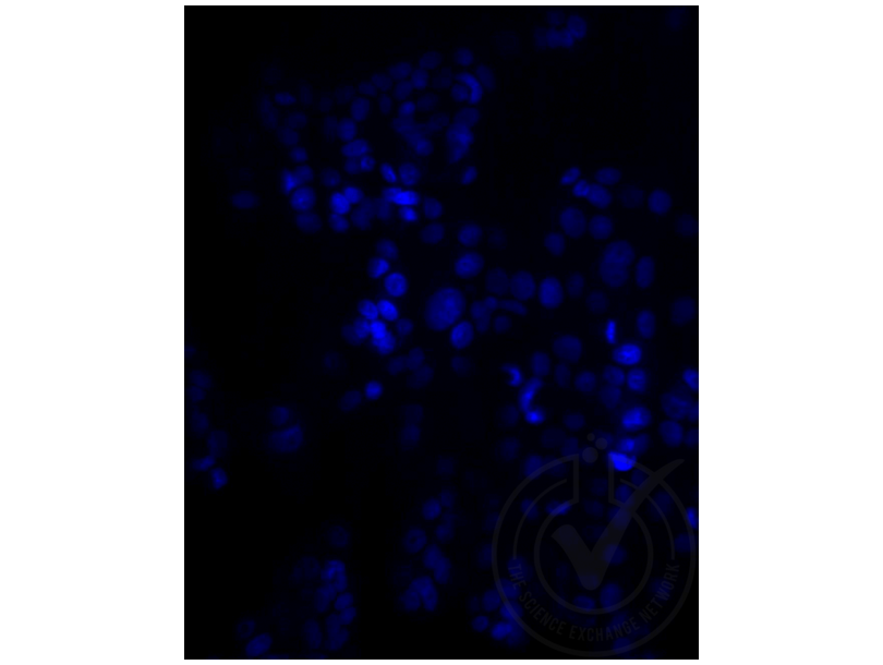
![Figure 3: HeLa cells stained with LRP2 (red) and a nuclear counterstain (blue).]() Figure 3: HeLa cells stained with LRP2 (red) and a nuclear counterstain (blue).
Figure 3: HeLa cells stained with LRP2 (red) and a nuclear counterstain (blue).
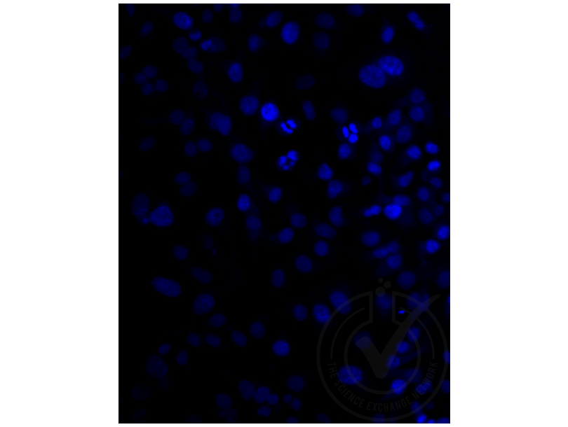
![Figure 4: MCF7 cells stained isotype control (red) and a nuclear counterstain (blue).]() Figure 4: MCF7 cells stained isotype control (red) and a nuclear counterstain (blue).
Figure 4: MCF7 cells stained isotype control (red) and a nuclear counterstain (blue).
![Figure 1: MCF7 cells with only a secondary antibody (red) and a nuclear counterstain (blue).]() Figure 1: MCF7 cells with only a secondary antibody (red) and a nuclear counterstain (blue).
Protocole
Figure 1: MCF7 cells with only a secondary antibody (red) and a nuclear counterstain (blue).
Protocole -
- Format
- Liquid
- Concentration
- 1 μg/μL
- Buffer
- Aqueous buffered solution containing 0.01M TBS ( pH 7.4) with 1 % BSA, 0.03 % Proclin300 and 50 % Glycerol.
- Agent conservateur
- ProClin
- Précaution d'utilisation
- This product contains ProClin: a POISONOUS AND HAZARDOUS SUBSTANCE, which should be handled by trained staff only.
- Stock
- -20 °C
- Stockage commentaire
- Store at -20°C. Aliquot into multiple vials to avoid repeated freeze-thaw cycles.
- Date de péremption
- 12 months
-
- Antigène
- LRP2 (Low Density Lipoprotein Receptor-Related Protein 2 (LRP2))
- Autre désignation
- Lrp2/Megalin (LRP2 Produits)
- Sujet
-
Synonyms: DBS, GP33, Low-density lipoprotein receptor-related protein 2, LRP-2, Glycoprotein 33, Megalin, LRP2
Background: Acts together with cubilin to mediate HDL endocytosis (By similarity). May participate in regulation of parathyroid-hormone and para-thyroid-hormone-related protein release.
- ID gène
- 4036
- UniProt
- P98164
- Pathways
- Metabolism of Steroid Hormones and Vitamin D, Thyroid Hormone Synthesis, Hormone Transport
-


